
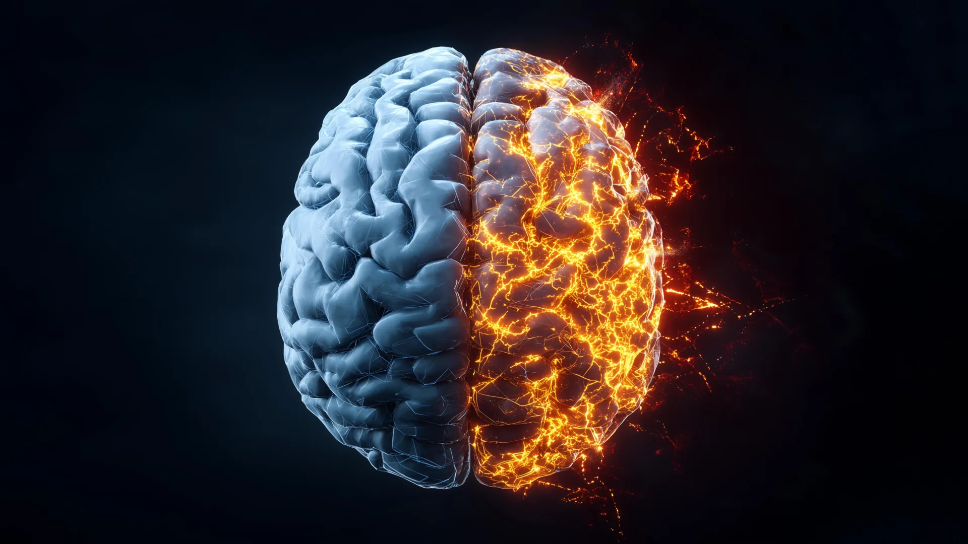
Researchers at Rice University have achieved a groundbreaking milestone in Alzheimer’s disease research, producing the first comprehensive, label-free molecular atlas of the Alzheimer’s brain in an animal model. This pioneering work, detailed in the prestigious journal ACS Applied Materials and Interfaces, offers an unprecedented and deeper understanding of the intricate ways in which the neurodegenerative disease initiates and progresses. Alzheimer’s disease, a relentless condition that claims more lives annually than breast and prostate cancers combined, presents a significant and growing public health challenge, underscoring the critical urgency of unraveling its underlying mechanisms.
Unlocking Brain Chemistry with Advanced Imaging
The innovative approach employed by the Rice University team combines an advanced light-based imaging methodology with sophisticated machine learning algorithms. This synergy allowed them to meticulously examine brain tissue from both healthy control animals and those exhibiting Alzheimer’s pathology. The results of their exhaustive analysis reveal a more complex picture than previously understood: the chemical changes intrinsically linked to Alzheimer’s disease are not confined to the well-known amyloid plaques. Instead, these alterations manifest throughout the entire brain, exhibiting uneven and remarkably complex spatial distribution patterns.
To achieve this level of detail in detecting subtle chemical shifts within brain tissue, the scientists harnessed the power of hyperspectral Raman imaging. This highly advanced form of Raman spectroscopy utilizes a focused laser beam to identify and map the unique chemical fingerprints of molecules present within the tissue. Each molecule possesses a distinct spectral signature, akin to a molecular barcode, which the laser can excite and detect.
Ziyang Wang, an electrical and computer engineering doctoral student at Rice and a first author on the study, elaborated on the significance of this technique. "Traditional Raman spectroscopy typically provides a single point measurement of chemical information at a specific molecular site," Wang explained. "Hyperspectral Raman imaging, however, revolutionizes this by repeating this measurement thousands of times across an entire tissue slice. This process effectively builds a comprehensive map, rendering a detailed picture that illustrates how chemical composition varies across different regions of the brain."
The research protocol involved scanning entire brains meticulously, slice by slice. Thousands of overlapping measurements were meticulously compiled to construct high-resolution molecular maps of both healthy and diseased brain tissue. A crucial aspect of this methodology is its "label-free" nature. This means the biological samples were not subjected to any pre-treatment with dyes, fluorescent proteins, or molecular tags, which can sometimes alter the natural state of the tissue or obscure subtle, endogenous changes.
"This label-free approach is paramount because it allows us to observe the brain precisely as it is, capturing a complete, unaltered portrait of its chemical makeup," Wang emphasized. "I firmly believe this makes the approach more unbiased and significantly better suited for discovering novel disease-related changes that might otherwise be overlooked or masked by introduced labels."
Machine Learning Deciphers Complex Alzheimer’s Damage Patterns
The sheer volume of data generated by the hyperspectral Raman imaging process was immense, necessitating the application of advanced machine learning (ML) techniques for analysis. The research team initially employed unsupervised ML algorithms. This approach allowed the algorithms to autonomously identify natural patterns and groupings within the complex chemical signals without any preconceived notions or prior assumptions about what to expect. These models effectively sorted and categorized the tissue based solely on its inherent molecular characteristics. Subsequently, the researchers utilized supervised ML. In this phase, they trained models to reliably distinguish between brain tissue exhibiting Alzheimer’s pathology and that of healthy controls. This crucial step enabled them to quantify the extent to which different brain regions reflected Alzheimer’s-related chemistry.
"Our analysis revealed a profound finding: the changes induced by Alzheimer’s disease are not uniformly distributed across the brain," Wang stated. "Instead, some regions exhibit substantial chemical alterations, while others remain comparatively less affected. This intricate, uneven pattern of damage could offer crucial insights into why Alzheimer’s symptoms manifest gradually over time and may also explain why therapeutic strategies that target only a single pathological pathway have historically met with limited success."
Unveiling Metabolic Disruptions in Critical Memory Regions
Beyond the accumulation of misfolded proteins, a hallmark of Alzheimer’s, the study identified broader metabolic differences between healthy and Alzheimer’s-affected brains. The levels of key biomolecules such as cholesterol and glycogen exhibited significant variations across different brain regions. The most pronounced contrasts were observed in areas critically responsible for memory formation and retrieval, particularly the hippocampus and the cerebral cortex.
Shengxi Huang, an associate professor of electrical and computer engineering and materials science and nanoengineering, and the corresponding author of the study, highlighted the functional significance of these findings. "Cholesterol plays a vital role in maintaining the structural integrity and function of brain cells, while glycogen serves as an essential local energy reserve," Professor Huang explained. "Taken together, these findings strongly support the evolving understanding that Alzheimer’s disease involves widespread disruptions in brain structure and energy balance, extending beyond just protein accumulation and misfolding." Professor Huang is also affiliated with several prominent research institutes at Rice, including the Ken Kennedy Institute, the Rice Advanced Materials Institute, and the Smalley-Curl Institute, underscoring the interdisciplinary nature of this research.
A Paradigm Shift in Understanding Alzheimer’s Progression
The genesis of this ambitious project stemmed from ongoing discussions among researchers seeking novel and more effective ways to study the complexities of the Alzheimer’s brain. Initially, the team’s focus was on analyzing relatively small areas of brain tissue. However, a conceptual leap led to the aspiration of mapping entire brains.
"At first, our scope was limited to measuring only small areas of brain tissue," Wang recalled. "Then, I began to ponder the possibility of mapping the entire brain to gain a much broader and more holistic view. It required several iterative rounds of rigorous testing and meticulous trial and error before the complex measurement and analysis techniques finally converged and began to work harmoniously together."
The moment the complete chemical map of the Alzheimer’s brain began to coalesce was met with immediate and significant impact. New and previously unseen patterns started to emerge that had been entirely invisible under conventional imaging modalities.
"Patterns emerged that had not been visible under regular imaging," Wang said, reflecting on the profound realization. "Witnessing those results unfold was deeply satisfying. It felt akin to revealing a hidden layer of information that had been present all along, simply awaiting the correct analytical approach to be unveiled."
By delivering the first detailed, dye-free chemical maps of the Alzheimer’s brain, this groundbreaking research offers a fundamentally more comprehensive and unbiased perspective on the disease’s progression. The research team expresses optimism that these findings will ultimately contribute to the development of earlier and more accurate diagnostic tools, as well as the formulation of more effective therapeutic strategies aimed at slowing or even halting the relentless progression of Alzheimer’s disease.
Broader Impact and Future Directions
The implications of this research extend far beyond the confines of the laboratory. The ability to visualize the chemical landscape of the brain in such detail, without the need for potentially confounding labels, opens up new avenues for research and clinical application.
Supporting Data and Context:
Alzheimer’s disease is the most common form of dementia, affecting millions worldwide. In the United States alone, it is estimated that over 6 million Americans are currently living with Alzheimer’s, a number projected to rise significantly in the coming decades. The economic burden of Alzheimer’s is also substantial, with the estimated cost of care in the U.S. reaching hundreds of billions of dollars annually. Traditional diagnostic methods often rely on cognitive assessments and imaging techniques that detect structural changes or amyloid plaques and tau tangles, which are typically indicative of later-stage disease. This new molecular atlas offers the potential to identify pathological changes at a much earlier, perhaps even preclinical, stage.
Timeline and Chronology:
While a precise start date for the project is not provided, the development of hyperspectral Raman imaging has been an ongoing area of scientific advancement over the past decade. The integration of machine learning for complex data analysis in biological imaging has also seen rapid progress in recent years. The publication in ACS Applied Materials and Interfaces, a journal known for its rigorous peer-review process, signifies the culmination of years of dedicated research, experimentation, and validation by the Rice University team. The study highlights a progression from smaller-scale analyses to the ambitious undertaking of mapping entire brain slices, suggesting a phased development of the methodology.
Potential Official Responses and Inferences:
While direct quotes from external parties are not available in the provided text, it is reasonable to infer that leading Alzheimer’s research foundations and government health agencies would view this work with significant interest. Organizations like the National Institute on Aging (NIA), a part of the National Institutes of Health (NIH), which partially funded this research, are constantly seeking innovative approaches to understanding and treating neurodegenerative diseases. The Alzheimer’s Association, a major non-profit organization dedicated to Alzheimer’s care, support, and research, would also likely recognize the potential impact of a label-free, comprehensive molecular atlas. Such advancements are crucial for accelerating the pace of drug discovery and developing more targeted interventions.
Fact-Based Analysis of Implications:
The "label-free" aspect of this technology is a critical differentiator. Many current molecular imaging techniques require the introduction of specific probes or labels to highlight particular molecules or structures. These labels, while useful, can sometimes interfere with cellular processes or introduce artifacts, potentially skewing results. By observing the brain’s natural chemical state, the Rice team has created a more authentic representation of disease pathology.
The discovery of uneven chemical changes and broader metabolic disruptions has significant implications for therapeutic development. Current Alzheimer’s treatments often focus on clearing amyloid-beta plaques or tau tangles. However, the uneven distribution of chemical alterations suggests that a single therapeutic target might not be sufficient to address the multifaceted nature of the disease. This research supports a paradigm shift towards more comprehensive, multi-target therapeutic strategies that consider the systemic metabolic and structural changes occurring in the brain.
Furthermore, the ability to map these changes at a molecular level could pave the way for the development of novel biomarkers for early diagnosis. By identifying specific chemical signatures associated with the earliest stages of Alzheimer’s, clinicians might be able to intervene before significant cognitive decline occurs, potentially improving patient outcomes dramatically.
The research was supported by significant funding from federal agencies and private foundations, including the National Science Foundation (grants 2246564 and 1934977), the National Institutes of Health (grant 1R01AG077016), and the Welch Foundation (grant C2144). This collaborative funding underscores the national and international recognition of the importance of Alzheimer’s research and the innovative nature of this project. The continued investment in such cutting-edge research is vital for making meaningful progress against this devastating disease.
