
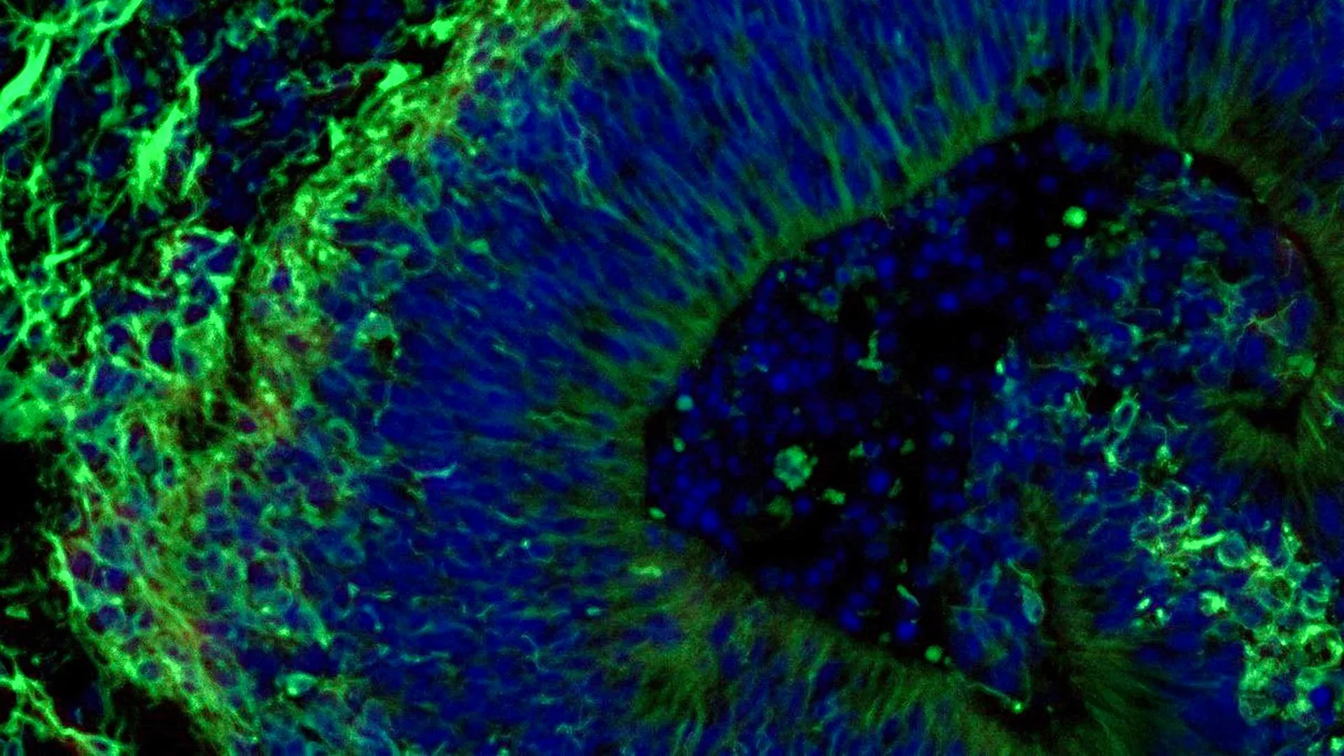
Scientists at Johns Hopkins Medicine have unveiled groundbreaking research utilizing miniature brain models derived from individuals with Alzheimer’s disease, offering a significant leap forward in predicting patient responses to medications targeting the condition’s challenging neuropsychiatric symptoms. This innovative approach, detailed in Alzheimer’s & Dementia: The Journal of the Alzheimer’s Association, suggests a future where Alzheimer’s treatment can be tailored to the individual, moving beyond the current one-size-fits-all approach.
Alzheimer’s disease, the most prevalent form of dementia, currently affects over 7 million Americans, with its debilitating impact extending beyond memory loss to encompass a range of neuropsychiatric symptoms such as anxiety, depression, and agitation. These symptoms are nearly universal among patients, yet the effectiveness of available treatments, primarily Selective Serotonin Reuptake Inhibitors (SSRIs), varies dramatically from person to person. This variability has long been a significant hurdle in providing optimal care.
The Johns Hopkins team’s research centers on brain organoids – small, lab-grown clusters of brain tissue that mimic key aspects of the human brain. By cultivating these organoids from the cells of individuals diagnosed with Alzheimer’s disease, researchers have created sophisticated models that recapitulate molecular hallmarks of the condition. This pioneering work not only deepens our understanding of Alzheimer’s progression but also lays the groundwork for developing more precise therapeutic strategies.
The Genesis of a Novel Approach: From Blood Cells to Brain Organoids
The study’s foundation was laid by collecting blood samples from individuals participating in the NIH-funded Johns Hopkins Alzheimer’s Disease Research Center. These samples, obtained with informed consent, were the starting point for a remarkable transformation. Through a process known as reprogramming, the researchers reverted the blood cells to a stem cell-like state, creating induced pluripotent stem cells (iPSCs). These iPSCs possess the remarkable ability to differentiate into virtually any cell type in the body, including the specialized neurons that form the brain.
The team then focused on developing hindbrain organoids. The hindbrain, located at the base of the brain, is crucial for regulating fundamental life functions such as breathing, sleep cycles, and heart rate. By guiding the iPSCs to self-organize, the researchers successfully generated pea-sized organoids that closely resembled the structure and cellular composition of the hindbrain. Crucially, these organoids contained neurons that produce serotonin, a key neurotransmitter implicated in mood regulation and a primary target of SSRI medications.
The scale of this endeavor is noteworthy. The study involved hundreds of organoids, each representing a unique individual with Alzheimer’s disease or a healthy control. This extensive collection represents one of the largest brain organoid studies conducted to date in the field of Alzheimer’s research, providing a robust platform for detailed molecular analysis.
Unveiling Alzheimer’s Molecular Signatures in Mini-Brains
Upon comparing organoids derived from individuals with Alzheimer’s disease to those from healthy counterparts, the Johns Hopkins researchers observed distinct molecular differences. These disparities were particularly evident in proteins involved in inter-neuronal communication, inflammatory processes, and pathways known to be dysregulated in Alzheimer’s pathology. These findings underscore the capacity of patient-derived organoids to accurately mirror the complex biological changes occurring in the brains of individuals with the disease.
Testing Therapeutic Efficacy: A Glimpse into Personalized Drug Response
The next critical phase of the research involved exposing these organoids to escitalopram oxalate, a widely prescribed SSRI antidepressant. The goal was to ascertain whether the organoids could predict how effectively this medication might alleviate neuropsychiatric symptoms associated with Alzheimer’s disease.
The results were compelling and offered a nuanced view of drug response. In a subset of organoids derived from Alzheimer’s patients, treatment with escitalopram oxalate led to an increase in proteins associated with serotonin signaling and enhanced communication between brain cells. These are precisely the pathways that SSRIs are designed to modulate. However, other organoids exhibited minimal to no molecular response to the medication.
"We used these organoids to model how some patients’ tissue may respond to a commonly prescribed SSRI," explained Dr. Vasiliki Machairaki, the study’s lead investigator and an associate professor of genetic medicine at Johns Hopkins University School of Medicine. "On a large-scale level, our model may eventually be used to identify subgroups of patients, based on underlying molecular mechanisms, who are more likely to respond to certain drugs and thus help us to create precise, targeted treatments in the long run."
This differential response observed in the organoids provides strong evidence for their potential in predicting individual patient outcomes. It suggests that by analyzing the molecular profile of a patient’s organoid, clinicians might be able to forecast their likely response to specific SSRIs, thereby avoiding trial-and-error prescribing and ensuring patients receive the most effective treatment from the outset.
Extracellular Vesicles: Tiny Messengers, Big Diagnostic Potential
Beyond cellular responses, the study also explored the role of extracellular vesicles (EVs). These are minuscule particles released by cells that carry vital molecular information, including proteins and RNA. The researchers hypothesized that EVs shed by brain organoids could serve as novel biomarkers for diagnosing Alzheimer’s disease, assessing its progression, and even predicting drug response.
Before and after administering escitalopram oxalate to the organoids, the scientists meticulously analyzed the proteins contained within the EVs released by both patient-derived and healthy control organoids. The findings revealed that these vesicles are rich in proteins essential for brain function, including those involved in neuronal communication, memory formation, and neurotransmitter release.
Notably, organoids from individuals with Alzheimer’s disease exhibited distinct alterations in several disease-associated proteins within their EVs. Specific proteins like RAB3A, NSF, and ATCAY, which play crucial roles in normal synaptic signaling, were found at lower levels in EVs from Alzheimer’s organoids.
Following escitalopram treatment, the levels of certain proteins within the EVs showed an increase, particularly in those connected to serotonin signaling and synaptic pathways targeted by antidepressants. Again, the degree of change varied significantly across different organoids, mirroring the varied cellular responses observed earlier.
"This variation raises the possibility that extracellular vesicles from brain organoids could eventually help identify which patients are most likely to benefit from a particular treatment," Dr. Machairaki stated. This opens up the exciting prospect of using EVs as a non-invasive diagnostic tool, potentially derived from a simple blood draw, to gain insights into a patient’s disease status and predict their therapeutic trajectory.
The Future of Alzheimer’s Care: Towards Precision Medicine
The implications of this research are profound, signaling a paradigm shift towards precision medicine in the management of Alzheimer’s disease. Currently, the journey to finding an effective treatment for the neuropsychiatric symptoms of Alzheimer’s can be long and arduous, involving numerous medication adjustments and potential side effects. The ability to predict drug response before administering a medication could revolutionize this process, saving patients considerable distress and optimizing their quality of life.
Dr. Machairaki and her team are already looking ahead, envisioning the development of more sophisticated organoid models. Their future research aims to incorporate immune cells and vascular-like networks into these organoids, creating even more realistic representations of living human brain tissue. This advancement could further enhance the accuracy and predictive power of these models.
The ultimate goal is for extracellular vesicles to function as a form of "liquid biopsy." Such a test, derived from a patient’s blood, could potentially offer a comprehensive picture of their Alzheimer’s disease, including an accurate diagnosis, an assessment of disease stage, and identification of specific disease subtypes that may respond best to particular therapeutic interventions.
While acknowledging that this study represents an early yet crucial step, the potential impact on millions of individuals affected by Alzheimer’s disease and their families is immense. By harnessing the power of patient-derived brain organoids and their secreted vesicles, scientists are forging a path towards a future where Alzheimer’s care is not only more effective but also deeply personalized.
The research was made possible through substantial funding from the National Institutes of Health (NIH) and the Paul G. Allen Frontiers Foundation, alongside support from the Richman Family Precision Medicine Center of Excellence in Alzheimer’s Disease at Johns Hopkins University. The collaborative effort included contributions from a multidisciplinary team of scientists from Johns Hopkins, Tymora Analytical Operations, and the University of Rochester School of Medicine and Dentistry.
The development of these advanced organoid models and the understanding of their secreted vesicles hold the promise of transforming the clinical landscape for Alzheimer’s disease. As research progresses, the prospect of truly personalized treatment plans, informed by a patient’s unique biological signature, moves closer to reality, offering a beacon of hope for improved outcomes and enhanced quality of life for those living with this devastating condition.

0 Comments