
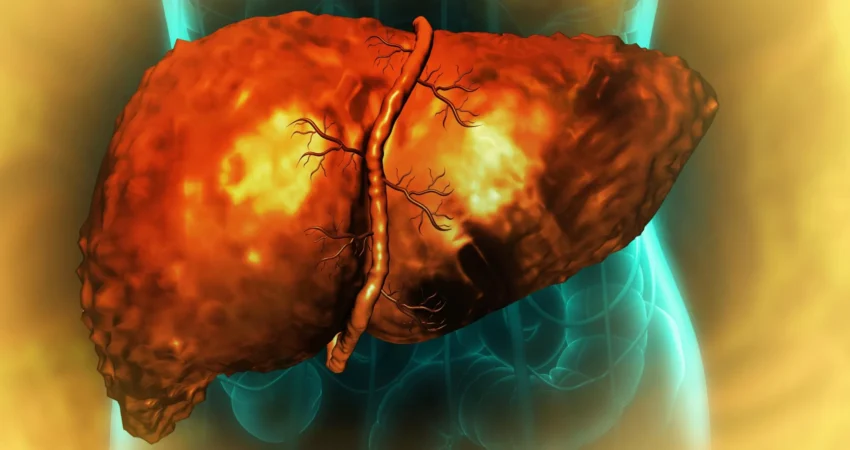
Excessive alcohol consumption can cripple one of the liver’s most extraordinary capacities: its innate ability to heal and regenerate after injury. Groundbreaking new research reveals that alcohol-induced damage can leave liver cells in a precarious limbo, an abnormal intermediate state from which they are unable to perform their essential functions or complete the vital process of regeneration, even after an individual ceases drinking. This cellular malfunction, previously a mystery, has now been linked to inflammation disrupting a fundamental cellular mechanism known as RNA splicing.
Researchers from the University of Illinois Urbana-Champaign, Duke University, and the Chan Zuckerberg Biohub Chicago have elucidated a critical pathway by which alcohol wreaks havoc on liver regeneration. Their findings, published in the prestigious journal Nature Communications, shed light on a molecular mechanism that could revolutionize the diagnosis and treatment of severe alcohol-associated liver disease, a condition that claims millions of lives globally each year.
The Liver’s Unparalleled Regenerative Power Under Siege
The liver stands apart from most human organs due to its remarkable capacity to regenerate. Following substantial damage, or even surgical removal of a portion, the remaining liver cells possess the ability to temporarily alter their identity, proliferate, and then mature back into fully functional tissue, thereby restoring lost mass and function. This intrinsic regenerative capability is crucial for maintaining the organ’s vital roles in metabolism, detoxification, and protein synthesis.
However, this extraordinary resilience is severely compromised in alcohol-associated liver disease (ALD), a progressive and often fatal condition that is the leading cause of liver-related mortality worldwide, contributing to an estimated 3 million deaths annually. The escalating burden of ALD underscores the urgent need to understand the underlying mechanisms of liver failure.
"For years, clinicians and researchers have observed that the liver’s ability to function and regenerate is critically impaired in patients suffering from alcohol-related hepatitis and cirrhosis," stated Auinash Kalsotra, a professor of biochemistry at the University of Illinois and a co-leader of the study. "Crucially, this impairment persists even after patients have abstained from alcohol. The absence of this understanding has left liver transplantation as the only viable life-saving option for individuals who reach the end-stage liver failure. Our investigation aimed to uncover the ‘why’ behind this failure, hoping to identify potential points for therapeutic intervention."
Kalsotra and his long-time collaborator, Anna Mae Diehl, a professor at Duke University School of Medicine, have dedicated years to unraveling the molecular intricacies that govern the liver’s regenerative processes. Their prior research had already established that during regeneration, mature liver cells undergo a temporary reprogramming, shifting their gene expression patterns to adopt characteristics of earlier developmental stages.
Cells Caught in a Molecular Stalemate
The initial stages of liver repair involve mature liver cells dedifferentiating, reverting towards a fetal-like progenitor state. These progenitor cells are less specialized and possess a heightened capacity for division, enabling them to generate new liver tissue. Following successful multiplication, these cells are expected to re-differentiate, returning to their mature, fully functional state to restore the organ’s integrity.
This established regenerative cycle became the focus of the researchers’ inquiry into what goes awry in the context of alcohol-associated liver disease. To investigate this, the team meticulously compared healthy human liver tissue samples with tissue obtained from individuals diagnosed with alcohol-related hepatitis or cirrhosis. The diseased samples were generously provided by Johns Hopkins University Hospital, a key partner in an initiative supported by the National Institute on Alcohol Abuse and Alcoholism (NIAAA), part of the National Institutes of Health (NIH).
A stark and consistent pattern quickly emerged from the analysis. In the diseased liver samples, cells had indeed initiated a transition away from their mature identity and begun to move towards a regenerative state. However, they failed to complete this critical journey. Instead, they became arrested, trapped in an intermediate, "quasi-progenitor" state, neither fully functional adult cells nor fully capable progenitor cells.
"These cells are essentially stuck in limbo," explained Ullas Chembazhi and Sushant Bangru, graduate students at the University of Illinois and co-first authors of the study. "Because they are not functioning, this places immense pressure on the remaining healthy cells. These healthy cells then attempt to regenerate, but they too often end up in this same unproductive quasi-progenitor state, ultimately leading to liver failure."
This cellular stalemate perpetuates a vicious cycle. As more and more cells succumb to this unproductive state, the pool of functional cells diminishes, compromising the liver’s ability to perform its essential tasks. The remaining healthy cells, facing an overburdened workload, attempt to compensate by initiating regeneration, inadvertently risking their own entrapment in the same debilitating condition.
RNA Splicing: The Unseen Culprit
To pinpoint the molecular defect preventing cells from completing their regenerative journey, the researchers delved into the intricate processes of protein production and RNA activity within liver cells. RNA molecules act as crucial intermediaries, transcribing genetic instructions from DNA into the cellular machinery responsible for building proteins. A fundamental step in this process, known as RNA splicing, involves the precise removal of non-coding regions and the joining of coding segments. This "editing" is critical because different combinations of spliced RNA segments can dictate the production of proteins with distinct functions or direct them to specific locations within the cell.
Moving beyond conventional studies that merely quantify RNA and protein levels, Kalsotra’s team employed advanced deep RNA sequencing and sophisticated computational analysis to meticulously examine the intricate patterns of RNA splicing.
"Our comparative analysis revealed a widespread and pervasive issue of RNA missplicing in alcohol-related liver disease, affecting thousands of genes," Kalsotra reported. "This missplicing had a profound impact on the functional capabilities of numerous proteins essential for liver health."
The sheer scale of the problem was significant. Missplicing was not an isolated event but a widespread phenomenon, potentially altering the function of a vast array of crucial proteins within damaged liver cells.
ESRP2 Deficiency: A Key Player in Splicing Errors
Delving deeper, the researchers identified a key factor contributing to these extensive splicing errors: a marked deficiency in a protein known as ESRP2 (Epithelial Splicing Regulatory Protein 2). ESRP2 plays a critical role in ensuring the accurate splicing of RNA molecules. In liver cells damaged by alcohol, the study found that ESRP2 levels were significantly reduced.
The consequences of this ESRP2 deficiency extended beyond the mere production of proteins. In many instances, the RNA errors introduced by the lack of proper splicing fundamentally altered the molecular instructions that dictate a protein’s cellular destination.
"Proteins must function at specific locations within the cell, and this localization is guided by sequences embedded within the protein itself," Kalsotra explained. "We discovered that in numerous cases, the very sequences responsible for directing a protein to its correct cellular compartment were misspliced. This is why our comprehensive analytical approach was so critical. We observed that while the quantity of RNA and protein might appear normal, the protein was not in the right place to perform its intended function. Due to missplicing, key proteins essential for effective liver regeneration were becoming trapped in the cytoplasm, when their proper function required them to be in the nucleus."
The nucleus houses the cell’s DNA and serves as the central control center for gene expression. The cytoplasm, the surrounding cellular environment, is where many other cellular processes take place. If proteins critical for regeneration remain sequestered in the cytoplasm instead of reaching the nucleus, they are effectively rendered inactive, despite their presence.
Evidence from Mouse Models Reinforces the ESRP2 Connection
To definitively establish whether the loss of ESRP2 directly contributes to the failure of liver regeneration, the researchers conducted experiments using mice genetically engineered to lack the gene responsible for producing ESRP2. These animal models exhibited patterns of liver injury and regenerative failure that closely mirrored the pathological conditions observed in human patients with advanced alcohol-related hepatitis.
This finding raised a critical subsequent question: What causes the reduction in ESRP2 levels in the first place? The research team traced the root cause back to inflammation.
When the liver processes alcohol, it incurs tissue damage, which in turn triggers an inflammatory response. This response attracts immune cells and specialized liver support cells to the injured areas. According to the study, these activated cells release a cascade of inflammatory and growth factors. The researchers discovered that these inflammatory signals actively suppress both the production and the functional activity of ESRP2.
Targeting Inflammation for Liver Repair
The team then moved to test a crucial hypothesis: could interrupting these inflammatory signals reverse the detrimental effects on RNA splicing and regeneration? In laboratory cultures of liver cells, they introduced a molecule designed to block the receptor for a key inflammation-promoting factor. Following this targeted intervention, ESRP2 levels showed a notable recovery, and the patterns of RNA splicing began to normalize.
This pivotal result strongly suggests that the inflammatory pathway could serve as a promising target for future therapeutic interventions. Instead of directly attempting to replace damaged liver tissue, novel treatments might focus on disrupting the inflammatory signals that impede the liver’s intrinsic regenerative capabilities.
Beyond therapeutic potential, the researchers also envision significant diagnostic applications. The presence of abnormally spliced RNA molecules could potentially serve as valuable biomarkers, aiding in the early identification and monitoring of alcohol-associated liver disease.
"I am genuinely hopeful that these findings will serve as a catalyst for future clinical studies," Kalsotra expressed. "We can potentially leverage these misspliced RNAs as diagnostic markers, or develop therapeutic strategies aimed at curbing the inflammation. If we can effectively correct these splicing defects, we may be able to significantly improve recovery outcomes and restore the health of damaged livers."
The research team’s comprehensive investigation was supported by substantial funding from the National Institutes of Health, the Chan-Zuckerberg Biohub Chicago, The Duke Endowment, and the Muscular Dystrophy Association. Specific grants from the NIH, including R01-AA010154, R01-HL126845, R21-HD104039, 5R01-DK077794, 1R56-DK1343340, and R24 AA025017, were instrumental in enabling this critical work. The collaborative effort involved scientists from the University of Illinois, Duke University, Johns Hopkins University School of Medicine, and Northwestern University, highlighting the power of interdisciplinary research in tackling complex health challenges.

0 Comments