
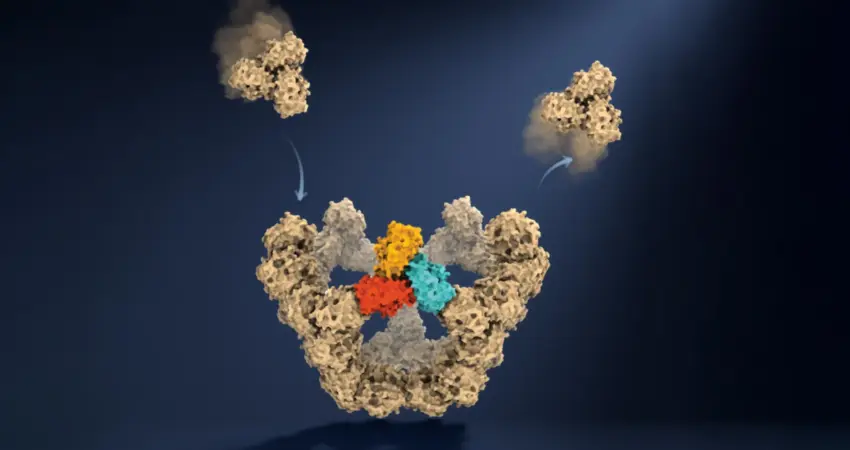
Researchers at the University of Oxford have achieved a groundbreaking feat in molecular biology, directly observing virus-like particles assemble themselves molecule by molecule. This pioneering work, published in the prestigious journal Nature, has illuminated the remarkably simple yet sophisticated rules governing the formation of complex biological structures with exceptional fidelity. For decades, scientists have grappled with understanding the intricate process of viral assembly, often relying on static snapshots or theoretical models. This new research provides an unprecedented, real-time view into how individual protein components spontaneously organize into precise architectural designs, a process critical for viral replication and a potential target for novel therapeutic interventions.
The Challenge of Observing Viral Assembly
Viruses, microscopic agents of infection, are essentially packages of genetic material encased within protective protein shells known as capsids. The size and complexity of these capsids vary enormously, with some composed of just a few dozen protein subunits, while others comprise thousands. A fundamental puzzle in virology has been how these individual protein building blocks, in the absence of any cellular machinery or external guidance, can reliably find their correct positions and assemble into the functional viral structure. This self-assembly process is not only essential for viral survival but also presents a fascinating case study in emergent order from simple components.
Previous attempts to unravel this mystery were hampered by the ephemeral nature of intermediate structures. During assembly, transient configurations of proteins form and dissolve, often existing for mere fractions of a second and in exceedingly low concentrations. This made them exceptionally difficult to detect and characterize using conventional observational techniques. Consequently, the scientific community’s understanding was largely built upon analyzing the initial raw materials (individual proteins) and the final, fully formed viral particles, leaving a significant gap in knowledge regarding the dynamic transitions between these states.
A Technological Leap: Mass Photometry Illuminates the Process
The Oxford team, led by Dr. Roi Asor, Dan Loewenthal, and Professor Philipp Kukura from the Department of Chemistry and the Kavli Institute for Nanoscience Discovery, overcame this observational hurdle by employing a cutting-edge technique called mass photometry. This method, developed at Oxford, quantifies the mass of individual molecules by precisely measuring the light they scatter. Crucially, the researchers developed an innovative method to confine individual molecules and maintain them under continuous observation, allowing for repeated measurements of their mass as the assembly process progressed.
"This method lets us study the assembly process directly; we just take a video!" exclaimed PhD student Dan Loewenthal, highlighting the revolutionary nature of their approach. By tracking the incremental addition of protein units and the resulting changes in mass, the scientists could effectively construct a molecular-level timeline of the assembly. This allowed them to witness, for the first time, the step-by-step construction of a virus-like particle.
The engineered virus-like particle used in the study comprised 60 identical protein units. This simplified yet representative model allowed for detailed analysis without the overwhelming complexity of natural viral systems. The ability to quantify the underlying molecular interactions and follow the growth of a single particle transformed the field, moving beyond theoretical reconstructions to empirical observation. "We can see the important intermediate structures appear and deduce a detailed model of the construction process," stated Professor Philipp Kukura, emphasizing the shift from inference to direct evidence.
Navigating the Molecular Maze: The Rules of Self-Assembly
The study revealed that viral self-assembly is akin to navigating a complex maze, where only specific pathways lead to successful completion. The protein components initially form weak, reversible connections. This inherent flexibility is a critical design feature, allowing the molecules to disassociate if they form an incorrect arrangement, thereby continuing to explore different combinations. This "trial and error" mechanism is fundamental to overcoming the sheer number of potential molecular interactions.
A key finding is the identification of crucial "molecular checkpoints" – stable intermediate structures that act as signposts, guiding the assembly process. The first of these significant milestones occurs when five larger protein building blocks coalesce to form a stable, closed pentagonal ring. This nucleation event is particularly challenging, requiring a specific alignment of components. However, once this initial stable structure is formed, the subsequent stages of assembly accelerate significantly. Each new stable configuration achieved requires fewer additional building blocks, allowing the particle to grow more rapidly towards its final form.
"A virus has to solve an extraordinary construction problem," explained Dr. Roi Asor. "Its components somehow have to find the right arrangement among a huge number of possibilities, without a blueprint or machinery directing the process. We can now watch what happens molecule by molecule and see the physical rules that make it possible."
The research highlights a crucial balance: the initial instability of weak bonds allows for correction of errors, preventing a single misplaced molecule from derailing the entire process. Once a sufficient number of components lock into the correct closed arrangement, the structure gains stability, creating a robust foundation for further growth. This combination of exploration through weak interactions and consolidation through stable structures ensures the remarkable reliability of viral assembly, even in the absence of explicit instructions.
Timeline of Discovery: A Decade of Pushing Boundaries
The insights gleaned from this Oxford study represent the culmination of years of dedicated research and technological development. While the fundamental concept of viral self-assembly has been recognized for decades, the ability to observe it in real-time is a more recent advancement.
- Early 2000s: Initial theoretical models and biochemical studies begin to outline the principles of protein self-assembly in viruses, focusing on the thermodynamics and kinetics of subunit interactions.
- Mid-2010s: Advances in microscopy and biophysical techniques allow for more detailed characterization of protein structures and their interactions, though direct observation of dynamic assembly remains elusive.
- Late 2010s: The development and refinement of mass photometry at Oxford provides a potential avenue for single-molecule observation and mass determination.
- Early 2020s: The Oxford team integrates mass photometry with novel confinement techniques, enabling the direct, real-time tracking of viral particle assembly.
- Publication in Nature: The findings are published, marking a significant leap in understanding the fundamental mechanisms of biological self-assembly.
Broader Implications: From Antivirals to Vaccines and Beyond
The ramifications of this research extend far beyond fundamental virology. A detailed understanding of how viral capsids are constructed offers unprecedented opportunities for developing new medical interventions.
Antiviral Drug Development:
By identifying the precise molecular interactions that drive viral assembly, scientists can now design drugs that specifically target and disrupt these critical steps. Interfering with the formation of the viral capsid could prevent the virus from encapsulating its genetic material or from maturing into an infectious particle. This could lead to a new class of antiviral therapies that are more targeted and potentially less prone to resistance development compared to existing treatments. For instance, compounds that mimic or block the formation of the initial pentagonal ring, a crucial nucleation event, could prove highly effective.
Vaccine Design:
The ability to engineer and understand self-assembling virus-like particles (VLPs) is also invaluable for vaccine development. VLPs, which mimic the structure of viruses but lack their genetic material and thus are non-infectious, are potent immunogens. They can be used to present viral antigens to the immune system, eliciting a robust protective response. The insights from this study could allow for the more efficient and precise design of VLPs for vaccines against a range of viral diseases, potentially leading to more effective and stable vaccine formulations.
Biotechnology and Nanotechnology:
The principles of self-assembly observed in viral systems are not unique to viruses. Self-assembly is a ubiquitous phenomenon in biology, responsible for the construction of cellular structures like cytoskeletons, organelles, and complex protein machinery. The Oxford team’s methodology offers a powerful new tool for studying these diverse biological processes. This could accelerate research in areas such as synthetic biology, where researchers aim to build novel biological systems from the ground up, and nanotechnology, where self-assembling molecular components are used to create new materials and devices.
Potential for New Materials:
Beyond medicine, the ability to control and engineer self-assembly at the molecular level could lead to the development of novel materials with unique properties. Imagine materials that can spontaneously form intricate structures, repair themselves, or adapt to their environment – principles inspired by biological self-assembly.
Expert Reactions and Future Directions
The scientific community has reacted with considerable enthusiasm to the Oxford team’s findings. Dr. Jack Tan from the MRC Weatherall Institute of Molecular Medicine and the CAMS-Oxford Institute, a co-author on the study, remarked, "The ability to understand these processes at the molecular level could have important applications in vaccine development and antivirals that disrupt viral assembly."
Professor Jane Davies, a leading virologist not involved in the study, commented, "This is a truly transformative piece of work. For years, we’ve been trying to infer these dynamics from static data. To actually see the assembly happen, molecule by molecule, is a game-changer. It validates many theoretical predictions and opens up entirely new avenues for therapeutic intervention."
Looking ahead, the Oxford researchers plan to apply their technique to study the assembly of more complex viral systems and to investigate how cellular factors might influence or regulate viral assembly in vivo. Understanding these in-cell dynamics will be the next frontier. Furthermore, the adaptability of mass photometry and confinement techniques suggests its broad applicability to studying the self-assembly of various macromolecular complexes, both biological and artificial. This research marks not just a significant advance in virology but a foundational step in our ability to understand and engineer the very building blocks of life.

0 Comments