
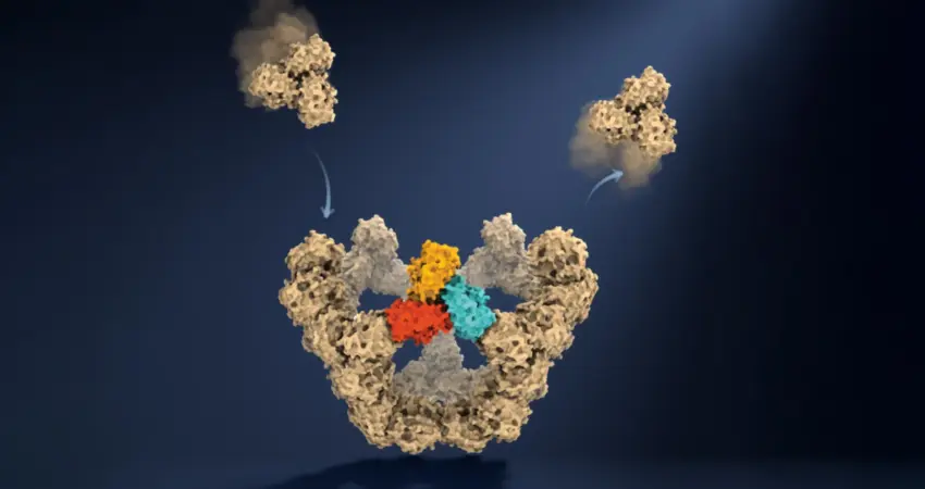
Researchers at the University of Oxford have achieved a significant breakthrough, directly observing for the first time the intricate process of virus-like particles assembling themselves, molecule by molecule. This pioneering work, published in the prestigious journal Nature, has unveiled the surprisingly simple yet remarkably robust rules governing the self-assembly of complex biological structures. For decades, scientists have grappled with directly visualizing this fundamental biological phenomenon, often relying on indirect evidence and theoretical models. This new research provides an unprecedented, real-time, molecular-level understanding of how individual protein components converge with remarkable precision to form the protective outer shells of viruses, known as capsids.
The implications of this discovery are far-reaching, offering a deeper insight into fundamental biological processes and opening new avenues for medical innovation. Understanding the precise mechanisms of viral capsid assembly is crucial for developing novel antiviral therapies that can disrupt this process, potentially rendering viruses incapable of replication. Furthermore, this knowledge can inform the design of more effective vaccines and advanced biotechnological tools. The findings address a long-standing scientific puzzle: how thousands of individual protein subunits, each with numerous potential connection points, consistently organize themselves into specific, functional three-dimensional structures, defying the statistical probability of random assembly.
The Molecular Maze: Navigating Assembly Pathways
The Oxford team likens the process of viral assembly to navigating a complex maze, where only specific pathways lead to a successful and stable outcome. Initially, the protein components form weak, transient interactions. This inherent flexibility is critical, allowing for a degree of "trial and error." If a particular arrangement of proteins proves unsuccessful or unstable, these weak bonds can easily break, releasing the components to explore alternative connections. This iterative process of forming and breaking bonds continues until a critical threshold of stable interactions is achieved.
Once a sufficient number of these protein building blocks lock into a correct, closed configuration, the resulting structure gains significant stability. These stable intermediate structures act as crucial "molecular checkpoints" along the assembly pathway. They effectively filter out the vast number of theoretically possible incorrect arrangements, funneling the process towards a limited set of highly probable and successful routes.
A pivotal moment in this assembly sequence is the formation of a closed pentagonal ring, comprised of five larger protein units. This initial stable structure is a critical milestone. Professor Philipp Kukura, a co-lead author from the Department of Chemistry and the Kavli Institute for Nanoscience Discovery at Oxford University, explained, "This is the first highly stable structure in the assembly process. Once it forms, each subsequent stable stage requires fewer additional building blocks, allowing the particle to grow more quickly." This cascading effect of stability accelerates the assembly process, ensuring efficient completion.
A Technological Leap: Witnessing Assembly in Real-Time
Capturing the ephemeral intermediate stages of viral self-assembly has historically posed a formidable challenge for researchers. Many of these structures exist for only fractions of a second and in minuscule quantities, making them nearly invisible to conventional observation techniques. Traditional methods typically allow scientists to examine the initial pool of raw materials and the final assembled particle, but the transformative journey between these two states remained largely obscured.
To surmount this hurdle, the Oxford researchers employed a sophisticated approach involving an engineered virus-like particle. This specific particle was constructed from 60 identical protein units, a common structural motif found in many viruses. The key to their success lay in the application of mass photometry, a cutting-edge technique developed at Oxford. Mass photometry quantifies the mass of individual molecules by meticulously measuring the light they scatter.
By integrating this powerful detection method with a novel experimental setup designed to confine individual molecules and maintain them under continuous observation, the team achieved an unprecedented level of detail. They were able to repeatedly measure the mass of a single, growing particle as additional protein subunits attached. "This method lets us study the assembly process directly, we just take a video!" exclaimed Dan Loewenthal, a PhD student and co-first author from the Department of Chemistry and the Kavli Institute for Nanoscience Discovery at Oxford. This "molecular movie" provided a direct, quantitative record of the assembly process, tracking the addition of each molecular building block over time.
Dr. Roi Asor, a co-lead author from the Department of Chemistry and the Kavli Institute for Nanoscience Discovery, elaborated on the significance of this technological advancement. "A virus has to solve an extraordinary construction problem," he stated. "Its components somehow have to find the right arrangement among a huge number of possibilities, without a blueprint or machinery directing the process. We can now watch what happens molecule by molecule and see the physical rules that make it possible." This direct observation contrasts sharply with previous reliance on snapshots or theoretical extrapolations.
The Elegance of Imperfection: A Robust Assembly Mechanism
The findings illuminate the ingenious strategy employed by these biological systems to overcome the overwhelming combinatorial complexity of assembly. Instead of attempting to explore every conceivable molecular arrangement, the protein components are guided through a finite sequence of stable intermediate states. These checkpoints ensure that the assembly process progresses efficiently and reliably towards the correct final structure.
This mechanism also sheds light on why the initial stage of assembly, known as nucleation, is often the most challenging. Once the first stable structure, such as the pentagonal ring, is formed, the subsequent growth phase accelerates significantly. This is because the stable intermediate structures provide a pre-defined framework that reduces the number of possible attachment sites for incoming subunits, streamlining the process.
A crucial element of this efficiency lies in the system’s tolerance for "mistakes." The weak initial interactions allow for incorrect pairings to readily dissociate. This "give" in the system provides ample opportunity for the protein building blocks to reorient and find more favorable connections. As Dr. Asor further explained, "The weak interactions give the system room to make mistakes. Most encounters don’t have to be successful: the components can separate and try again. But once enough of them come together in the right closed arrangement, the structure becomes stable, and assembly can move forward. That combination of trial and error followed by locking in successful structures is what makes the process so reliable." This delicate balance between transient flexibility and robust stability is the hallmark of this elegant self-assembly mechanism.
Implications for Medicine and Beyond
The ability to meticulously observe and understand the molecular interactions that govern viral capsid assembly holds immense promise for the development of novel therapeutic strategies. By identifying key steps or weak points in the assembly pathway, scientists may be able to design drugs that specifically inhibit these processes, thereby preventing the formation of functional viruses. This could lead to a new generation of antiviral medications with precisely targeted mechanisms of action.
Furthermore, these insights can be leveraged to enhance existing medical interventions. For instance, understanding how viral proteins self-assemble can aid in the design of more immunogenic and stable virus-like particle (VLP) vaccines. VLPs mimic the structure of viruses but are non-infectious, making them excellent platforms for eliciting protective immune responses. Dr. Jack Tan from the MRC Weatherall Institute of Molecular Medicine and the CAMS-Oxford Institute, a co-author on the study, highlighted this potential: "The ability to understand these processes at the molecular level could have important applications in vaccine development and antivirals that disrupt viral assembly."
The impact of this research extends beyond the realm of virology. Self-assembly is a fundamental principle that underpins a vast array of biological structures and processes. From the intricate scaffolding within cells to the formation of complex protein machinery involved in cellular functions, self-assembly is a ubiquitous and essential phenomenon. The Oxford team’s innovative approach, which allows for the observation of these intricate construction processes at the single-molecule level, offers a powerful new tool for investigating the origins of biological complexity. By providing a direct window into how simple interactions give rise to sophisticated biological architectures, this research paves the way for future investigations into a wide spectrum of biological phenomena.
A Glimpse into the Future of Biological Research
The successful observation of virus-like particle self-assembly molecule by molecule represents a significant leap forward in our ability to study fundamental biological processes. The integration of mass photometry with novel confinement techniques has unlocked a previously inaccessible realm of molecular dynamics. This technological advancement not only validates long-held theoretical models but also provides a rich dataset for refining our understanding of the physical principles governing biological self-organization.
The potential for this methodology to be applied to other self-assembling biological systems is immense. Researchers can now envision studying the assembly of protein filaments, the formation of cellular organelles, and the intricate mechanisms of protein folding with unprecedented clarity. This opens up exciting possibilities for drug discovery, biomaterials science, and fundamental biological research.
The University of Oxford has a long-standing tradition of pioneering research in the life sciences and nanotechnology. This latest publication in Nature further solidifies its position at the forefront of molecular imaging and biophysics. The collaborative efforts of chemists and biologists, leveraging cutting-edge instrumentation, have demonstrated the power of interdisciplinary research in tackling complex scientific challenges. The findings are expected to stimulate further investigation into the rules of self-assembly across the biological sciences, potentially leading to paradigm shifts in our understanding of life’s fundamental building blocks and their remarkable ability to construct ordered complexity from seemingly simple beginnings.

0 Comments