
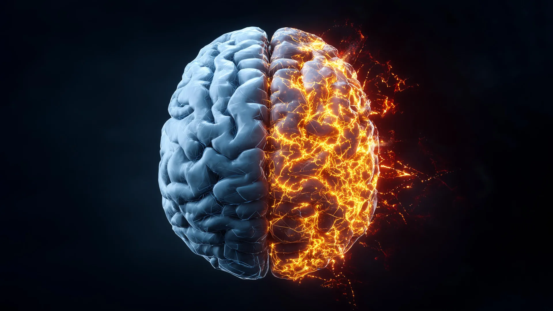
Researchers at Rice University have achieved a significant breakthrough in Alzheimer’s disease research, generating the first comprehensive, label-free molecular atlas of the Alzheimer’s brain within an animal model. This pioneering work provides an unprecedentedly detailed view of how the devastating neurodegenerative disease initiates and progresses, offering critical insights into the complex biochemical landscape of affected brain tissue. Alzheimer’s disease, a relentless condition that claims more lives annually than breast and prostate cancers combined, underscores the profound urgency to unravel its underlying mechanisms and develop effective interventions.
The multidisciplinary team employed a sophisticated combination of advanced light-based imaging and cutting-edge machine learning algorithms to meticulously examine brain tissue from both healthy and Alzheimer’s-affected animal subjects. Their findings, recently published in the esteemed scientific journal ACS Applied Materials and Interfaces, reveal a startling reality: the chemical alterations associated with Alzheimer’s disease are not confined to the well-known amyloid plaques. Instead, these profound changes manifest throughout the brain in intricate, uneven, and complex patterns, suggesting a more diffuse and multifaceted pathology than previously understood.
Illuminating Brain Chemistry with Advanced Laser Imaging
To precisely detect these subtle yet critical chemical shifts, the scientists leveraged hyperspectral Raman imaging, a highly advanced form of Raman spectroscopy. This powerful technique utilizes a laser to meticulously scan tissue, identifying the unique "chemical fingerprints" of individual molecules.
"Traditional Raman spectroscopy typically provides a single data point of chemical information for a specific molecular site," explained Ziyang Wang, an electrical and computer engineering doctoral student at Rice and a first author on the study. "Hyperspectral Raman imaging, in contrast, repeats this measurement process thousands of times across an entire tissue slice. This iterative approach enables us to construct a complete molecular map, revealing with remarkable clarity how chemical composition varies across different anatomical regions of the brain."
The research team meticulously scanned entire brain samples, section by section, accumulating thousands of overlapping measurements. This exhaustive process allowed them to assemble high-resolution molecular maps of both healthy and diseased brain tissue. A key advantage of this methodology is its label-free nature, meaning the samples were analyzed without the introduction of dyes, fluorescent proteins, or molecular tags that could potentially alter the native state of the tissue.
"This absence of labeling is crucial," Wang emphasized. "It means we observed the brain in its natural state, capturing a complete and unaltered portrait of its chemical makeup. We believe this unbiased approach is more conducive to discovering novel disease-related changes that might otherwise be overlooked with traditional labeling methods."
Machine Learning Deciphers Uneven Alzheimer’s Damage Patterns
The sheer volume of data generated by the hyperspectral Raman imaging process presented a significant analytical challenge. To overcome this, the researchers turned to the power of machine learning (ML). Initially, they employed unsupervised ML algorithms. This approach allowed the algorithms to identify inherent patterns within the vast array of chemical signals without any preconceived notions or prior assumptions about the data. The models independently sorted tissue samples based purely on their molecular characteristics. Subsequently, the team utilized supervised ML, training specific models to accurately distinguish between Alzheimer’s-affected and non-Alzheimer’s samples. This crucial step helped quantify the degree to which different brain regions exhibited Alzheimer’s-related chemistry.
"Our analysis revealed a profound insight: the chemical changes induced by Alzheimer’s disease are not uniformly distributed throughout the brain," stated Wang. "Certain brain regions displayed significant chemical alterations, while others appeared comparatively less affected. This uneven distribution pattern offers a compelling explanation for why Alzheimer’s symptoms often emerge gradually and why therapeutic strategies that target only a single pathological aspect have historically met with limited success."
Metabolic Disruption in Critical Memory Centers
Beyond the detection of protein aggregation, a hallmark of Alzheimer’s, the study uncovered broader metabolic differences between healthy and diseased brains. The researchers observed significant variations in the levels of cholesterol and glycogen across different brain regions. These discrepancies were most pronounced in areas vital for memory function, specifically the hippocampus and the cortex, structures critically involved in learning and memory consolidation.
"Cholesterol plays an indispensable role in maintaining the structural integrity of brain cells, while glycogen serves as a readily accessible local energy reserve," explained Shengxi Huang, an associate professor of electrical and computer engineering and materials science and nanoengineering at Rice, and the corresponding author of the study. "Taken together, these findings strongly support the hypothesis that Alzheimer’s disease involves widespread disruptions in brain structure and energy metabolism, extending beyond the mere accumulation and misfolding of proteins." Professor Huang is also affiliated with several prominent Rice research centers, including the Ken Kennedy Institute, the Rice Advanced Materials Institute, and the Smalley-Curl Institute, highlighting the collaborative nature of this research.
A Paradigm Shift in Understanding Alzheimer’s Progression
The genesis of this ambitious project can be traced back to ongoing discussions among researchers seeking novel methodologies to study the complex pathology of the Alzheimer’s brain.
"Initially, our investigations were limited to analyzing only small, localized areas of brain tissue," recalled Wang. "Then, a pivotal question arose: what if we could map the entire brain to gain a significantly broader and more holistic perspective? It required considerable experimentation and iterative refinement of both measurement techniques and analytical approaches before we achieved a harmonious integration of imaging and data processing."
The moment the complete chemical map of the Alzheimer’s brain began to coalesce, the impact was immediate and transformative.
"Patterns began to emerge that had simply been invisible with conventional imaging techniques," Wang remarked. "Witnessing those results was profoundly satisfying. It felt akin to uncovering a hidden layer of information that had been present all along, awaiting the right analytical tools to be revealed."
By providing the first detailed, dye-free chemical maps of the Alzheimer’s brain, this groundbreaking research offers a significantly more comprehensive understanding of the disease’s multifaceted nature. The research team expresses optimism that these findings will pave the way for earlier and more accurate diagnosis of Alzheimer’s disease and, crucially, support the development of more effective strategies to slow its relentless progression.
The research was generously supported by grants from the National Science Foundation (awards 2246564 and 1934977), the National Institutes of Health (grant 1R01AG077016), and the Welch Foundation (award C2144), underscoring the significant national and institutional investment in tackling this critical public health challenge. The implications of this work extend beyond fundamental scientific understanding, offering a glimmer of hope for improved patient outcomes in the future.
