
Scientists at Johns Hopkins Medicine have unveiled groundbreaking research that could revolutionize the way Alzheimer’s disease is treated, offering a personalized approach to managing the condition’s complex neuropsychiatric symptoms. The study, published in the esteemed Alzheimer’s & Dementia: The Journal of the Alzheimer’s Association, details the development and use of miniature, patient-derived brain organoids that demonstrate a remarkable ability to predict individual responses to medications commonly used to alleviate symptoms such as anxiety, depression, and agitation. This pioneering work not only deepens our understanding of Alzheimer’s disease mechanisms but also paves the way for more precise and effective therapeutic strategies.
A Leap Forward in Personalized Alzheimer’s Care
Alzheimer’s disease, the most prevalent form of dementia, affects an estimated 7 million Americans, posing a significant and growing public health challenge. While a cure remains elusive, managing the profound neuropsychiatric symptoms that plague nearly all patients is crucial for improving their quality of life. Selective serotonin reuptake inhibitors (SSRIs) are frequently prescribed for these symptoms, yet their efficacy varies dramatically among individuals. This variability has long been a source of frustration for clinicians and patients alike.
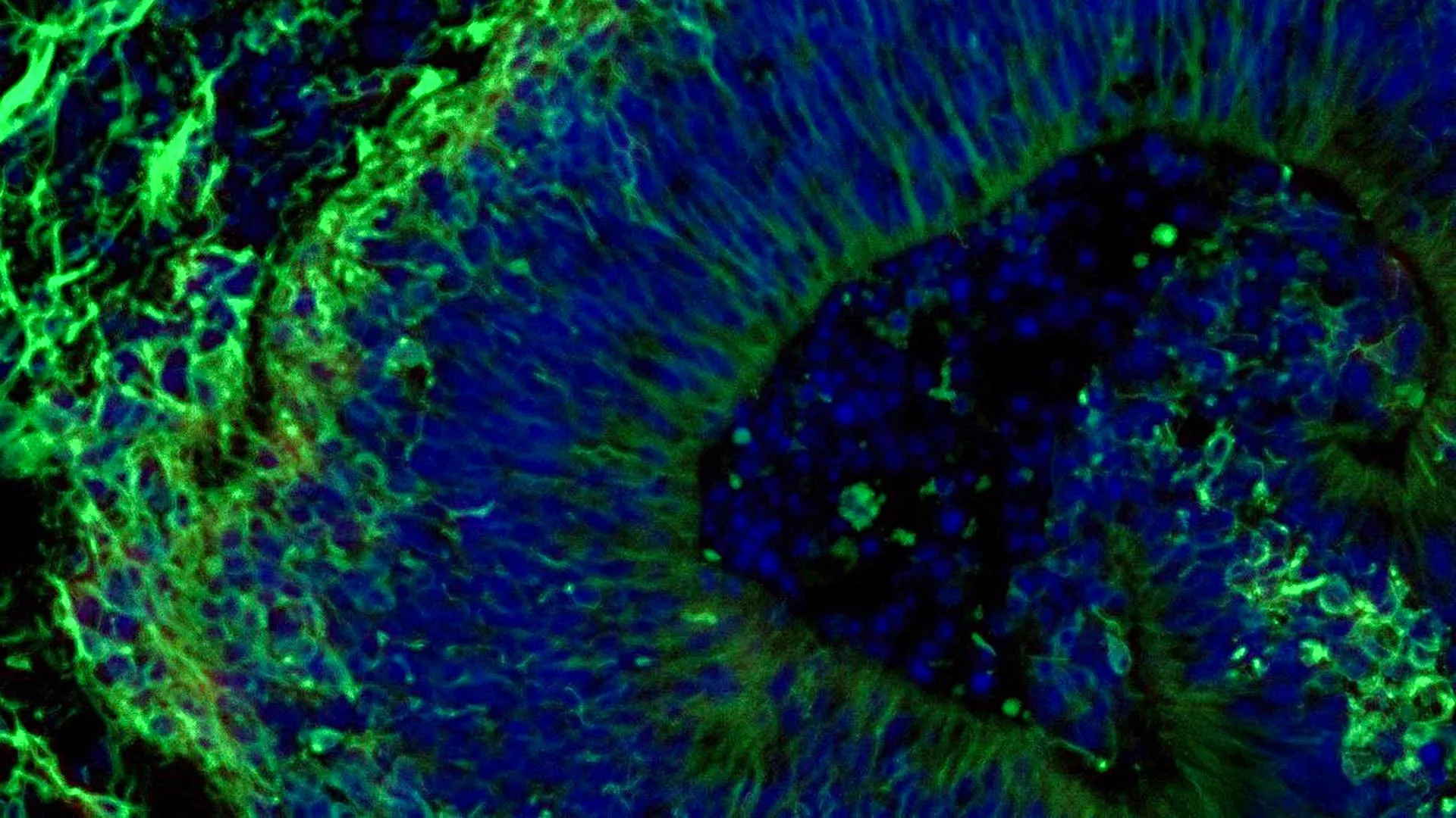
The Johns Hopkins team, led by Dr. Vasiliki Machairaki, an associate professor of genetic medicine, has developed a novel system using laboratory-grown brain tissues, known as organoids, derived from the cells of people with Alzheimer’s disease. These "mini-brains" are designed to mimic specific regions of the human brain, in this case, the hindbrain, which plays a vital role in regulating fundamental life functions such as breathing, sleep, and heart rate. By observing how these organoids react to specific medications, researchers can gain unprecedented insights into how individual patients might respond.
"Our study suggests that large-scale, patient-derived brain organoids and the vesicles they secrete can help us stage Alzheimer’s disease, investigate the mechanisms that drive it and assess how patient subgroups may respond to different treatments," stated Dr. Machairaki. "This is a critical step towards realizing the promise of personalized medicine for Alzheimer’s disease."
The Genesis of the Mini-Brains: From Blood Cells to Brain Tissue
The innovative research began with a meticulous collection of blood samples from individuals diagnosed with Alzheimer’s disease, obtained with ethical approval from the NIH-funded Johns Hopkins Alzheimer’s Disease Research Center. These samples provided the foundational material for creating the organoids.
The researchers employed a sophisticated reprogramming technique to transform ordinary blood cells back into induced pluripotent stem cells (iPSCs). These remarkable iPSCs possess the inherent ability to differentiate into virtually any cell type in the body. By utilizing iPSCs sourced from both individuals with Alzheimer’s disease and healthy control subjects, the team was able to cultivate hindbrain organoids. These organoids were engineered to contain specialized brain cells, or neurons, specifically those that produce the crucial neurotransmitter serotonin, a key target for many antidepressant medications.
Through a carefully controlled process, these reprogrammed cells were encouraged to self-organize into small, pea-sized clusters that closely resemble the structure and cellular composition of the hindbrain. The study’s scope was significant, involving hundreds of organoids meticulously representing individual Alzheimer’s patients alongside those from healthy participants. Dr. Machairaki believes this represents one of the most extensive brain organoid studies conducted to date in the field of Alzheimer’s research, underscoring the depth and breadth of the investigation.
Unveiling Molecular Signatures of Alzheimer’s in Organoids
A pivotal aspect of the research involved observing the distinct molecular characteristics that emerged within the patient-derived organoids. Compared to organoids grown from the cells of healthy individuals, those cultivated from Alzheimer’s patients exhibited notable differences in key proteins. These discrepancies were particularly evident in proteins involved in intercellular communication within the brain, inflammatory pathways, and other molecular cascades known to be associated with the progression of Alzheimer’s disease. These findings provide compelling evidence that the organoids effectively recapitulate critical pathological hallmarks of the disease at a cellular and molecular level.
The next critical phase of the study involved treating these organoids with escitalopram oxalate, a widely prescribed SSRI antidepressant. The results were illuminating. In a subset of organoids derived from Alzheimer’s patients, the medication demonstrably enhanced the levels of proteins involved in serotonin signaling and improved communication between brain cells. These are precisely the pathways that SSRIs are designed to modulate. However, a significant observation was that other organoids derived from different patients showed minimal or no discernible molecular response to the same medication.
"We used these organoids to model how some patients’ tissue may respond to a commonly prescribed SSRI," Dr. Machairaki explained. "On a large-scale level, our model may eventually be used to identify subgroups of patients, based on underlying molecular mechanisms, who are more likely to respond to certain drugs and thus help us to create precise, targeted treatments in the long run." This observation directly addresses the challenge of differential drug response and offers a potential solution through personalized predictive testing.
Extracellular Vesicles: Tiny Messengers of Disease and Drug Response
Beyond observing changes within the organoids themselves, the research team delved into the role of extracellular vesicles (EVs). These minuscule particles, secreted by cells, act as potent carriers of molecular information, including proteins, RNA, and lipids. The study explored whether EVs released by the brain organoids could serve as valuable biomarkers for diagnosing Alzheimer’s disease, gauging its progression, or even predicting a patient’s response to treatment.
The scientists meticulously collected EVs from both the Alzheimer’s patient-derived organoids and the healthy control organoids, both before and after treatment with escitalopram. Analysis of the proteins contained within these EVs revealed critical insights. The EVs were found to carry proteins integral to essential brain functions, such as neuronal communication, memory formation, and neurotransmitter release.
Significantly, EVs originating from the Alzheimer’s organoids displayed clear alterations in several disease-associated proteins. Notably, the levels of RAB3A, NSF, and ATCAY – proteins crucial for the normal signaling processes between brain cells – were found to be reduced in the Alzheimer’s organoids’ EVs. This reduction suggests a disruption in synaptic function, a hallmark of neurodegenerative diseases.
Furthermore, following escitalopram treatment, the levels of certain proteins within the EVs shifted. These changes were particularly pronounced in proteins linked to serotonin signaling and synaptic pathways that are the primary targets of antidepressant medications. The observed variation in these molecular responses across different organoids, mirroring the individual patient responses, reinforced the potential of EVs as predictive tools.
"Some organoids displayed strong molecular responses, while others showed little or no change," Dr. Machairaki noted. "This variation raises the possibility that extracellular vesicles from brain organoids could eventually help identify which patients are most likely to benefit from a particular treatment." This could translate into a future where a simple test of EVs could guide clinicians in selecting the most effective medication for a given patient, minimizing trial-and-error and improving therapeutic outcomes.
The Future of Brain Organoids: Towards a "Liquid Biopsy"
The implications of this research are far-reaching. The ability to accurately predict drug response could lead to a paradigm shift in Alzheimer’s treatment, moving away from a one-size-fits-all approach towards highly individualized care plans. This could not only enhance the effectiveness of existing medications but also accelerate the development of new therapies by enabling researchers to identify patient subgroups that are most likely to benefit from novel drug candidates during clinical trials.
Looking ahead, Dr. Machairaki and her team are committed to advancing the sophistication of their brain organoid models. Their future work includes incorporating immune cells and vascular-like networks that mimic the intricate structure of blood vessels. The inclusion of these components is expected to make the organoids even more representative of living human brain tissue, thereby increasing their predictive power and translational relevance.
The ultimate vision is for these extracellular vesicles to serve as a "liquid biopsy." This non-invasive test could potentially revolutionize Alzheimer’s diagnosis, allowing for earlier detection, accurate staging of the disease, and even the identification of specific disease subtypes. Such a diagnostic tool would be invaluable for both clinical management and the pursuit of targeted therapeutic interventions.
"With additional research, she hopes extracellular vesicles might one day function as a type of liquid biopsy," the study concluded. "Such a test could potentially help diagnose Alzheimer’s disease, determine its stage, and identify a patient’s particular disease subtype." This represents a significant step towards a future where Alzheimer’s disease can be diagnosed and treated with unprecedented precision.
The study received substantial support from the National Institutes of Health (NIH) through various grants, including T32 AG058527, R01AG052510, P30AG066507, 1RF1AG083801, AGR01054771, AGR01050515, AGR01046543, and AGR01071522. Additional funding was provided by the Paul G. Allen Frontiers Foundation and the Richman Family Precision Medicine Center of Excellence in Alzheimer’s Disease at The Johns Hopkins University. The collaborative effort involved a multidisciplinary team, including researchers from Johns Hopkins, Tymora Analytical Operations, and the University of Rochester School of Medicine and Dentistry, underscoring the collaborative nature of modern scientific discovery.
While this research represents an early but significant stride, its potential to transform the landscape of Alzheimer’s disease care is undeniable, offering a beacon of hope for millions affected by this devastating condition. The ongoing development of these sophisticated organoid models promises to unlock deeper insights into the complexities of Alzheimer’s and accelerate the delivery of truly personalized and effective treatments.

0 Comments