
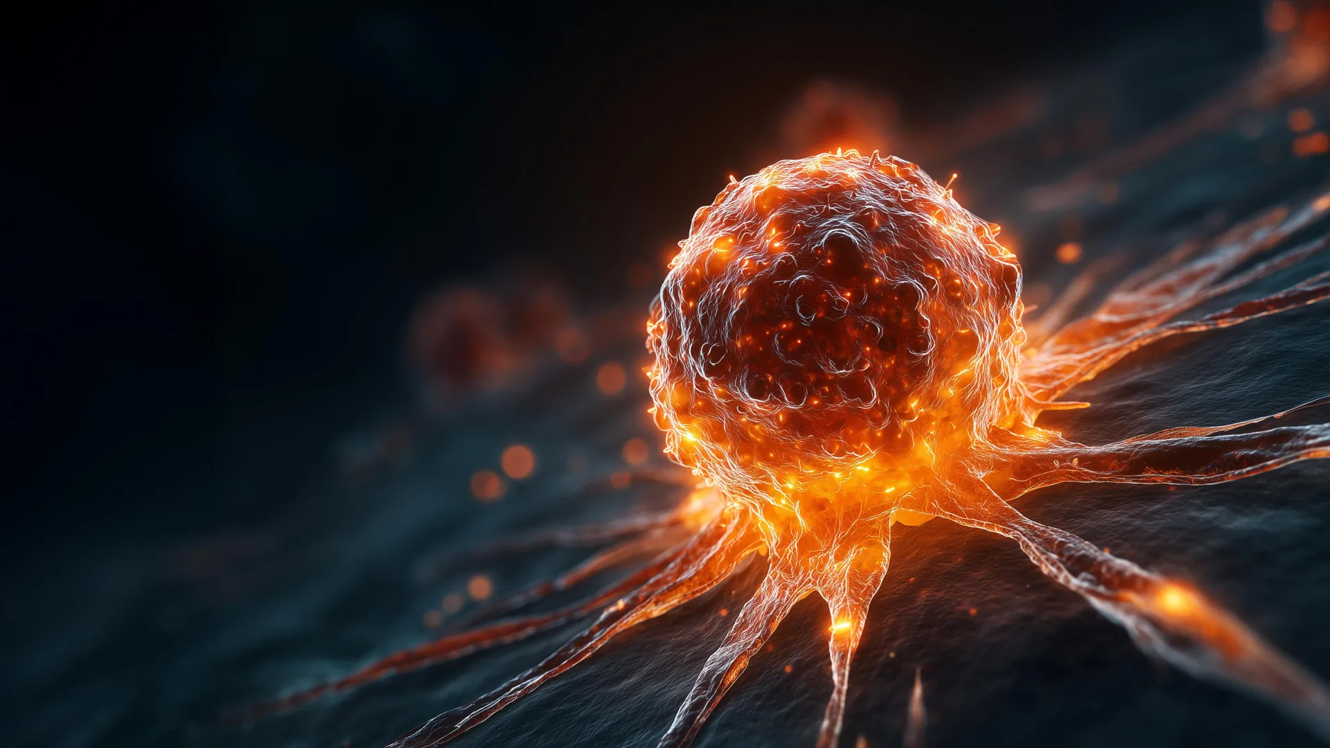
Researchers at the University of Missouri are pioneering a groundbreaking method to precisely identify cancer patients who stand to gain the most from targeted therapies. This innovative approach involves illuminating tumors within medical scans, offering a more accurate and efficient diagnostic pathway. At the heart of this development is a novel antibody designed to act as a beacon, guiding physicians toward the most effective treatment strategies.
Illuminating Cancer’s Presence: A Molecular "Flashlight" for Precision Medicine
The key to this advancement lies in the ingenious work of Barry Edwards, an associate professor of biochemistry at the University of Missouri School of Medicine. Professor Edwards has engineered a remarkably small antibody, a molecular key designed to specifically seek out and bind to EphA2. This protein is known to be significantly overexpressed in a wide array of human cancers, making it a promising target for both diagnostic and therapeutic interventions.
The ingenuity of Edwards’ design doesn’t stop at creating a selective antibody. He has further enhanced its utility by attaching a radioactive marker. This marker, when administered to a patient, allows the antibody to become visible during a positron emission tomography (PET) scan. This creates what can be aptly described as a molecular "flashlight," capable of casting a distinct glow on tumors that exhibit high concentrations of EphA2.
Experiments conducted on laboratory mice have provided compelling evidence of this technique’s efficacy. Professor Edwards’ research has demonstrated that the EphA2-targeting minibody clearly delineates tumors expressing the protein, distinguishing them from surrounding healthy tissues. These preclinical results suggest a significant potential for this tagged antibody to revolutionize cancer detection. It could enable physicians to pinpoint cancers harboring EphA2 and, critically, to identify patients who are most likely to respond favorably to targeted therapies. Such therapies are specifically designed to attack EphA2-positive tumor cells while sparing healthy tissue, a crucial distinction for minimizing side effects and maximizing treatment efficacy.
"By discerning which patients exhibit high or low levels of EphA2, we can precisely determine their likelihood of benefiting from a targeted cancer treatment," Professor Edwards explained. His dual appointment in the College of Arts and Science further underscores the interdisciplinary nature of his research. "There is a clear ethical and practical imperative to avoid administering treatments that are unlikely to be effective for a particular patient. This novel process we have developed has the potential to significantly reduce wasted time and resources, thereby accelerating the progress of precision medicine."
A Leap Forward: Speed, Invasiveness, and Diagnostic Insight
The current standard of care for evaluating tumors in cancer patients typically relies on invasive biopsies and Magnetic Resonance Imaging (MRI) scans. While these methods have been foundational in oncology, they are not without their limitations. Biopsies, by their very nature, involve surgical procedures, carrying inherent risks and discomfort for patients. Furthermore, both biopsies and MRIs can be time-consuming, often requiring days or even weeks to yield definitive results. Crucially, they frequently offer only a limited view of the specific molecular landscape within cancer cells, particularly the expression levels of key proteins like EphA2.
Professor Edwards’ work, conducted using advanced imaging technology at the University of Missouri’s Molecular Imaging and Theranostics Center, aims to overcome these challenges. His vision is to transition this promising technique from its current preclinical stage to human clinical trials within the next seven years. This ambitious timeline reflects the urgent need for more efficient and patient-friendly diagnostic tools in oncology.
"This new targeted approach offers a significant advantage by being noninvasive," Professor Edwards emphasized. "Furthermore, the diagnostic insights can be obtained within hours, a stark contrast to the days often required by traditional methods. For patients who may be traveling long distances to seek specialized treatment, this speed can be an enormous benefit." The ability to streamline the diagnostic process, making it both easier and faster for both patients and clinicians, represents a significant stride towards achieving the goals of precision medicine. This "win-win" scenario, as Professor Edwards described it, promises to enhance the patient experience and optimize clinical decision-making.
Background and Context: The Rise of Targeted Therapies and EphA2
The development of targeted cancer therapies represents a paradigm shift in oncology. Unlike traditional chemotherapy, which broadly affects rapidly dividing cells (both cancerous and healthy), targeted therapies are designed to interfere with specific molecules or pathways that are crucial for cancer cell growth and survival. This specificity is intended to lead to more effective treatments with fewer debilitating side effects.
The identification of specific biomarkers, such as proteins like EphA2, is fundamental to the success of targeted therapies. The Eph family of receptor tyrosine kinases and their ligands, the ephrins, play crucial roles in cell-to-cell adhesion, cell migration, and tissue patterning during embryonic development. However, their dysregulation is frequently observed in various cancers, contributing to tumor growth, invasion, and metastasis.
EphA2, in particular, has emerged as a significant oncogenic driver. Its overexpression is associated with more aggressive tumor phenotypes, poorer prognosis, and resistance to certain conventional therapies across a spectrum of cancers, including breast, ovarian, lung, and pancreatic cancers. This widespread involvement makes EphA2 a highly attractive target for therapeutic intervention. Several therapeutic strategies are currently being explored or are already in clinical use that target EphA2, including monoclonal antibodies, small molecule inhibitors, and antibody-drug conjugates.
The challenge, however, has been accurately and efficiently identifying which patients’ tumors express sufficient levels of EphA2 to warrant these targeted treatments. This is where Professor Edwards’ immunoPET agent comes into play. By providing a direct, noninvasive visualization of EphA2 expression, it acts as a crucial bridge between the availability of targeted therapies and their optimal application.
Supporting Data and Preclinical Evidence
The study, titled "Preclinical evaluation of anti-EphA2 minibody-based immunoPET agent as a diagnostic tool for cancer," published in the esteemed journal Molecular Imaging and Biology, details the rigorous preclinical validation of this technology. While specific quantitative data from the mouse models are proprietary to the research institution and not fully disclosed in the initial report, the qualitative findings are significant.
The research team employed various cancer cell lines and xenograft models in mice. These models were chosen to represent different cancer types and varying levels of EphA2 expression. Following the administration of the radiolabeled anti-EphA2 minibody, PET scans were performed. The resulting images clearly demonstrated a strong correlation between the signal intensity detected by the PET scanner and the known EphA2 expression levels in the implanted tumors. Tumors with high EphA2 expression showed robust radiotracer uptake, appearing as brightly lit areas on the PET scans. Conversely, tumors with lower or absent EphA2 expression exhibited significantly diminished or no detectable signal.
Histopathological analysis of the tumor tissues post-imaging further confirmed the specificity of the minibody’s binding. The distribution of the radiolabeled antibody within the tumor microenvironment was consistent with the known cellular localization of EphA2. This meticulous cross-validation between imaging data and biological analysis provides strong support for the accuracy and reliability of the diagnostic tool.
Broader Impact and Implications for Precision Oncology
The implications of this research extend far beyond improved diagnostic accuracy. The development of this EphA2-specific immunoPET agent has the potential to significantly impact several facets of cancer care:
Enhanced Patient Selection for Targeted Therapies
The most immediate benefit is the ability to precisely select patients who are most likely to respond to EphA2-targeted treatments. This avoids exposing patients to therapies that will not be effective, thereby preventing unnecessary side effects, financial burdens, and delays in receiving potentially life-saving alternative treatments.
Early Detection and Characterization
While primarily developed as a tool for treatment selection, the high sensitivity of PET imaging, combined with the specificity of the antibody, could also contribute to earlier detection and more accurate characterization of certain cancers. Identifying EphA2-positive lesions at an earlier stage could open up opportunities for timely intervention.
Monitoring Treatment Response
In the future, this technology might also be explored for monitoring treatment response. Changes in EphA2 expression or tumor avidity for the radiolabeled minibody could potentially indicate whether a targeted therapy is effectively controlling the cancer.
Advancing Theranostics
This work is a prime example of theranostics – a field that combines diagnostic and therapeutic capabilities. By visualizing the target (EphA2) with a diagnostic agent, clinicians can then confidently administer a therapeutic agent that targets the same molecule. This integrated approach maximizes treatment precision.
Reducing Healthcare Costs
By avoiding ineffective treatments and potentially shortening diagnostic timelines, this technology has the potential to contribute to significant cost savings within the healthcare system. More efficient patient selection and quicker diagnostic pathways can optimize resource allocation.
Accelerating Drug Development
For pharmaceutical companies developing EphA2-targeted therapies, this diagnostic tool could streamline clinical trials. It would allow for more efficient patient recruitment based on biomarker status, potentially leading to faster approval and availability of new drugs.
Future Directions and Timeline
Professor Edwards’ team is actively engaged in refining the anti-EphA2 minibody and the associated radiolabeling process. The current focus is on optimizing the stability of the radiotracer and ensuring its safety and efficacy in more complex preclinical models. The ambitious goal of initiating human clinical trials within seven years requires continued research, regulatory approvals, and potential collaborations with pharmaceutical partners.
The journey from preclinical research to clinical application is often lengthy and complex, involving multiple phases of human trials to assess safety, efficacy, and optimal dosing. However, the strong preclinical data and the clear clinical need for such a diagnostic tool provide a solid foundation for optimism.
The University of Missouri’s commitment to fostering innovation in medical research, exemplified by investments in facilities like the Molecular Imaging and Theranostics Center, plays a vital role in enabling breakthroughs like this. As the field of precision medicine continues to evolve, tools that enable highly specific patient stratification and treatment selection will become increasingly indispensable. Professor Edwards’ work on the EphA2-targeting immunoPET agent stands as a testament to the power of molecular engineering and advanced imaging in transforming cancer care.
