
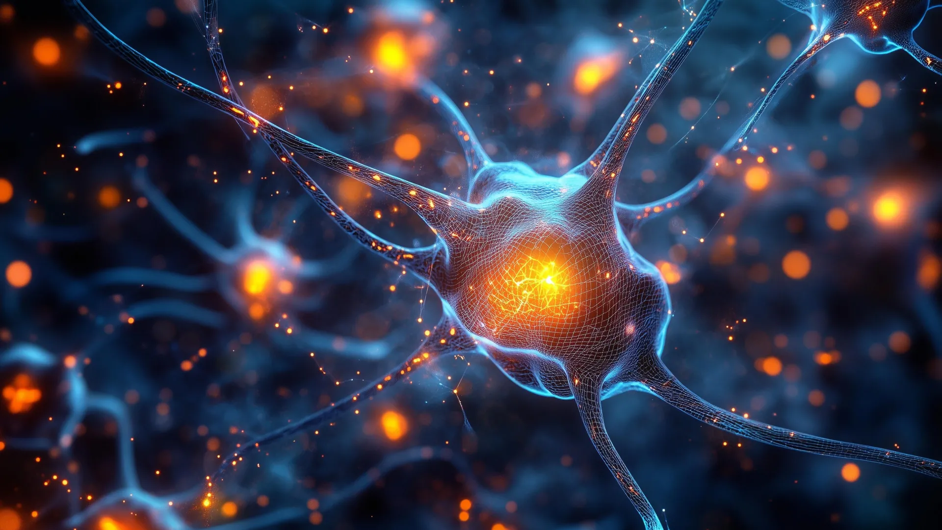
Scientists have identified a group of neurons located in an ancient region of the brain that plays a key role in helping animals focus. These cells appear to improve attention by filtering out distractions and directing the brain toward the most important information.
Unlocking the Secrets of Selective Attention: An Ancient Brainstem Circuit Revealed
BALTIMORE, MD – In a groundbreaking discovery that could reshape our understanding of attention and lead to novel treatments for attention-related disorders, researchers at Johns Hopkins University have pinpointed a network of neurons in an evolutionarily ancient part of the brainstem that is critical for an animal’s ability to focus. These newly identified cells act as a sophisticated filter, enabling the brain to discern and prioritize crucial information while effectively suppressing distracting stimuli. This finding challenges long-held assumptions about the neural underpinnings of attention, suggesting that a fundamental mechanism for focus predates the development of more complex brain structures like the prefrontal cortex.
The research, conducted in mice and recently published in the esteemed journal Nature Communications, has been lauded with an editorial highlight, underscoring its significance. The implications of this discovery are far-reaching, as the identified brainstem circuit is believed to be conserved across the entire vertebrate lineage, meaning it is present in species ranging from fish and birds to humans. This shared evolutionary heritage opens up exciting avenues for investigating attention deficits in humans, such as Attention-Deficit/Hyperactivity Disorder (ADHD) and autism spectrum disorder.
Challenging Prefrontal Cortex Dominance: A Look Back in Evolution
For decades, the scientific community largely attributed the sophisticated control of selective spatial attention – the ability to focus on specific stimuli in a complex environment while ignoring others – primarily to the prefrontal cortex. This region of the brain, which is significantly more developed in primates, including humans, is responsible for higher-level cognitive functions such as planning, decision-making, and working memory. However, this prevailing view presented a significant evolutionary puzzle: how do animals with less developed prefrontal cortices, such as birds and fish, exhibit remarkable abilities in focusing their attention?
"If we really go back in evolution, for hundreds of millions of years, birds have had this ability, fish have had this ability. And they do not typically have a highly developed prefrontal cortex, so how does the brain solve this problem?" queried lead author Ninad Kothari, a postdoctoral fellow in Johns Hopkins’ Department of Psychological and Brain Sciences. "We were able to identify an evolutionarily old region in the brainstem which affords this ability."
This persistent evolutionary question propelled the Johns Hopkins team to explore alternative neural substrates for attention. Their investigation led them to the brainstem, a foundational part of the central nervous system that controls basic life functions and serves as a crucial relay station for sensory and motor information.
The Brainstem’s "Focus Filter": Identifying the Key Neurons
The researchers’ meticulous work revealed a network of inhibitory neurons residing within the brainstem that play an indispensable role in attentional filtering. These neurons, present across a vast spectrum of vertebrate species, were the focus of the team’s experimental design. The initial impetus for investigating these specific brainstem cells stemmed from earlier studies conducted by senior author Shreesh Mysore, a neuroscientist specializing in neural circuits and behavior, and his colleagues, which explored attentional mechanisms in non-mammalian vertebrates like birds, frogs, and turtles.
To rigorously test the function of these brainstem neurons, the research team devised a sophisticated visual attention task for their mouse models. The task was designed to mimic the challenges humans face when distinguishing important signals from noise. Mice were presented with visual cues on a screen, with rewards contingent upon their correct response to stimuli appearing directly in their field of vision, while simultaneously being presented with distracting cues positioned at the periphery.
The initial phase of the experiment saw the mice performing the task with considerable accuracy, demonstrating their capacity for selective attention. However, a pivotal moment occurred when the researchers employed optogenetic techniques to temporarily deactivate these specific brainstem neurons. The results were immediate and striking.
"When we inactivate these neurons, the mice become hyper distractable," Kothari stated. The animals’ performance on the attention task plummeted, indicating a severe impairment in their ability to filter out irrelevant information and concentrate on the target stimuli.
Empirical Evidence: Disabling Neurons Leads to Increased Distractibility
To ensure that the observed deficits were not attributable to secondary factors such as visual impairments or motor coordination issues, the scientists conducted a series of supplementary experiments. These control tests meticulously ruled out any confounding variables, confirming that the mice’s decline in performance was specifically linked to their attentional processing capabilities.
The core finding was unequivocal: the inactivation of these brainstem neurons directly resulted in the loss of the animals’ ability to effectively weigh competing sensory inputs and prioritize the most relevant signal. The mice struggled to differentiate between the crucial target information and the surrounding distractions.
"The only thing impaired was their ability to take the competing pieces of information, compare them, and pay attention to the location with the most important information," explained Mysore. He further elaborated on the critical role of these neurons, likening this part of the brain to an "attentional selection engine." This engine, he posited, is fundamental to answering the crucial question: "’What is most important information I should pay attention to right now?’"
Broader Implications: A New Frontier for Treating Attention Disorders
The discovery of this ancient brainstem circuit’s role in attention opens up a significant new avenue for understanding and potentially treating attention-related disorders in humans. Conditions such as ADHD and autism spectrum disorder are characterized by difficulties in selective attention, where individuals may struggle to filter out distractions or to focus on relevant information.
The Johns Hopkins team is now eager to delve deeper into the precise mechanisms by which these brainstem neurons modulate spatial attention across the diverse range of vertebrate species. A key area of future research will be to ascertain whether these neurons perform a comparable function in humans.
"All the evidence to date suggests that these neurons exist in humans too," Mysore affirmed, expressing optimism about the potential relevance of their findings to human neurobiology. "But are they responsible for selective spatial attention in humans? An exciting hypothesis is that they play a crucial role."
The research team envisions future studies that will investigate the activity patterns of these brainstem neurons in individuals diagnosed with ADHD and autism. If these cells are found to function differently in these populations, it could pave the way for the development of more precise and effective therapeutic interventions. This could include targeted pharmacological treatments or behavioral therapies designed to modulate the activity of this fundamental attentional circuit, offering hope for improved outcomes for millions affected by these conditions.
The study’s authors also include Arunima Banerjee, Qingcheng (Jessica) Zhang, and Wen-Kai You from Johns Hopkins University, whose contributions were integral to this significant scientific advancement. The federally funded research signifies a crucial step forward in deciphering the complex neural architecture that underpins our ability to navigate and interact with the world around us.
A Chronology of Discovery: From Evolutionary Puzzles to Neural Circuits
The journey to this discovery likely began with long-standing observations in comparative neurobiology and ethology, the study of animal behavior in natural environments. For years, researchers noted the sophisticated attentional capabilities of animals with seemingly simpler brains, creating an evolutionary paradox that the prefrontal cortex-centric model struggled to explain.
Mid-20th Century Onwards: Early comparative studies in neuroscience begin to highlight the functional similarities in behavior across diverse vertebrate species, even when brain structures differ significantly. The ability of birds to locate specific prey in visually complex environments, or fish to evade predators in schooling formations, hints at underlying attentional mechanisms that are not solely reliant on highly evolved cortical structures.
Late 20th – Early 21st Century: Advancements in neuroimaging techniques and molecular biology allow for more detailed investigation into the neural circuits of various animal models. Researchers like Shreesh Mysore begin to focus on more evolutionarily conserved brain regions, including the brainstem, in their quest to understand fundamental cognitive processes. Studies on birds, amphibians, and reptiles, known for their less developed prefrontal cortices compared to mammals, likely provide crucial early clues about the involvement of subcortical structures in attention.
Circa 2010s: The development and refinement of optogenetic tools, which allow for precise control of neural activity using light, become instrumental. These tools enable researchers to selectively activate or inhibit specific neuron populations in living animals. This technological leap is critical for testing the causal role of identified neuronal groups.
Early 2020s: The Johns Hopkins University team, building on years of foundational research and leveraging advanced techniques, focuses on a specific network of inhibitory neurons within the brainstem. Through carefully designed behavioral experiments in mice, they systematically manipulate the activity of these neurons.
Mid-2023 (Publication Period): The experimental results strongly indicate that these brainstem neurons are essential for filtering distractions and enabling focused attention. The study is conducted, analyzed, and prepared for publication.
Recent Publication in Nature Communications: The findings are published in a leading scientific journal, receiving significant attention from the research community and being highlighted editorially. This marks the formal announcement of the discovery.
Present Day and Beyond: The scientific community begins to digest the implications of the findings. Further research is initiated to explore the precise molecular mechanisms, the functional parallels in humans, and the potential therapeutic applications for attention disorders.
Supporting Data and Future Research Directions
While the article does not present raw numerical data, the description of experimental outcomes provides strong qualitative evidence. The "success" of mice in the attention task prior to neuronal inactivation, followed by a significant decline when the neurons are silenced, serves as direct evidence of their functional importance. The successful ruling out of vision and movement problems further strengthens the conclusion that the impairment is specific to attentional processing.
Future research will likely involve:
- Detailed Electrophysiological Recordings: Measuring the electrical activity of these brainstem neurons during attention tasks in awake, behaving animals to understand their firing patterns and responses to stimuli.
- Molecular and Genetic Profiling: Identifying the specific neurotransmitters and receptors involved in the function of these neurons, which could reveal drug targets.
- Comparative Studies Across Vertebrates: Investigating the precise location and connectivity of these neurons in a wider range of species, including primates, to confirm their evolutionary conservation and functional equivalence.
- Human Studies: Developing non-invasive methods, such as fMRI or EEG, to study the activity of analogous brainstem regions in humans, particularly in individuals with ADHD and autism.
- Intervention Studies: Exploring whether stimulating or modulating these neurons can improve attentional performance in animal models of attention disorders.
The identification of this ancient brainstem circuit represents a significant leap in our understanding of attention, offering a new perspective on a fundamental cognitive ability that is essential for learning, survival, and social interaction.

0 Comments