
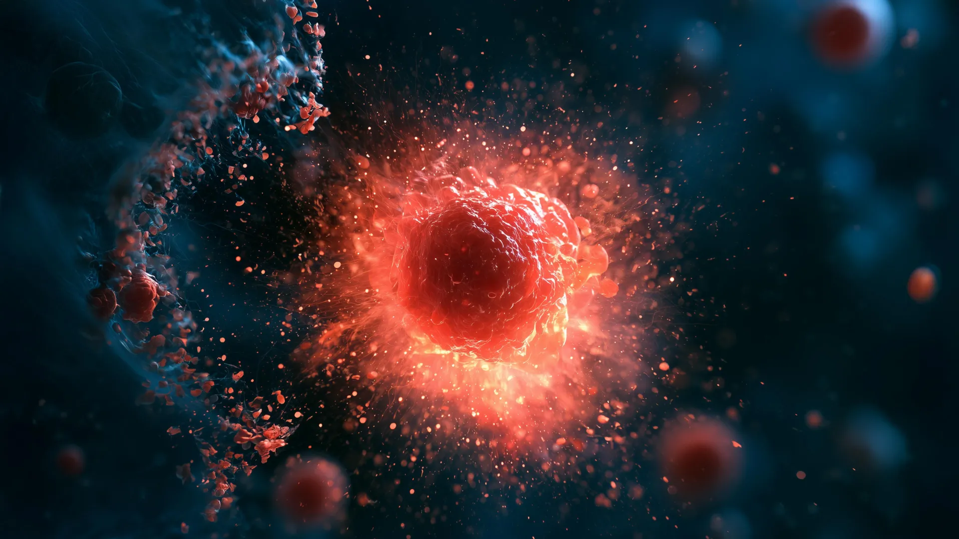
Stanford University researchers have identified a remarkable new type of immune cell, dubbed "ruptoblasts," that annihilates nearby cells by undergoing a rapid, explosive self-destruction. This dramatic process is so swift and complete that the cell effectively vanishes within minutes, leaving virtually no cellular debris behind. The groundbreaking discovery, made in the regenerative marvels of planarian flatworms, opens new avenues for understanding ancient immune strategies and could potentially inform future medical interventions.
The Enigmatic Power of Planarian Regeneration
The discovery of ruptoblasts was made in planarian flatworms, a class of simple aquatic animals celebrated for their extraordinary regenerative capabilities. These creatures possess an astonishing ability to regrow entire bodies from even tiny fragments. This remarkable resilience, honed over hundreds of millions of years of evolution, makes them invaluable subjects for scientists seeking to unravel the secrets of tissue repair and immune defense. Studying how these ancient organisms have maintained and adapted their immune systems could offer profound insights applicable to modern medicine, particularly in areas like wound healing, cancer therapy, and organ transplantation.
The research, detailed in a study published in the prestigious journal Cell, introduced the term "ruptoblasts" to describe these newly identified cells, a name derived from their explosive response triggered by a specific hormone. "We never expected that a cell could just explode like a bomb and kill the cells surrounding it," remarked senior author Bo Wang, an associate professor of bioengineering at Stanford’s Schools of Engineering and Medicine. This sentiment underscores the unexpected nature of the finding, which challenges conventional understandings of cellular defense mechanisms.
Unraveling the Mystery of Foreign Tissue Rejection
The initial observation of ruptoblasts stemmed from an investigation into a long-standing question in flatworm biology: their capacity to differentiate between self and non-self tissues. Chew Chai, a postdoctoral researcher in Dr. Wang’s laboratory, was at the forefront of this inquiry. To explore this question, Chai conducted an experiment that, in essence, created "Frankenstein" flatworms. He meticulously sliced individual worms lengthwise and then fused each with a segment from a different, unrelated worm.
While flatworms are renowned for their robust self-repair, these artificially created composite organisms exhibited a distinct rejection response towards the foreign tissue. This reaction bore a striking resemblance to the way a human immune system might reject a transplanted organ, a process driven by complex immunological recognition and defense. However, the cellular mechanisms underlying this rejection in flatworms proved to be profoundly different from anything observed in human immunology.
"It’s this huge inflammatory response," explained Chai, who is the lead author of the paper. "Like there’s a fire and an alarm goes off, and the cells just blow up." This vivid description highlights the immediate and dramatic nature of the flatworms’ immune reaction.
Activin: The Hormone Triggering Explosive Defense
Previous research into flatworm regeneration had already established the crucial role of the hormone activin in their survival. This hormone exhibits a delicate balance in flatworms: elevated levels can impair their ability to regenerate lost tissues, while low levels can interfere with their reproductive capabilities.
As the fused "Frankenstein" worms began the process of rejecting the foreign tissue, Chai observed a significant surge in activin levels. This hormonal spike was followed by a period of chronic inflammation. While the animals did not succumb immediately, they ultimately perished within several days of the fusion. Further experiments revealed that simply injecting activin into healthy, non-fused flatworms induced a comparable inflammatory response, solidifying its role as a key trigger for this extreme defensive reaction.
To delve deeper into the cellular events occurring during this inflammatory cascade, Chai employed advanced techniques. Live cell microscopy allowed for real-time observation of cellular behavior, while flow cytometry, a sophisticated method utilizing lasers to analyze and sort cells, provided quantitative data. By labeling cells with different fluorescent dyes, Chai could track and isolate those that responded to activin.
The Genesis of "Ruptosis" and the Ruptoblast
Through these meticulous observations, a small population of cells was identified that exhibited an astonishing behavior: they suddenly burst open, releasing potent substances that annihilated neighboring cells before disappearing entirely within a mere five minutes. Chai and Wang christened this unique and rapid cell death process "ruptosis," and the cells responsible for it, "ruptoblasts."
Unprecedented Speed and Efficiency in Cell Death
The defining characteristic of ruptosis is its unparalleled speed and thoroughness compared to other known forms of programmed cell death. "Some mammalian cells and bacteria may also do an explosive sort of cell death, but the timescale is really long. They are exploding, but it’s more like pores that slowly leak things out over the course of several hours," Chai elaborated. "Ruptosis happens within seconds to minutes." This stark contrast in timing highlights the exceptional nature of ruptoblast activity.
This rapid destruction effectively transforms each ruptoblast into a highly localized and potent weapon. Instead of a gradual release of harmful agents, the cell unleashes its destructive payload almost instantaneously. This concentrated burst of activity suggests a highly efficient and targeted mechanism for eliminating threats.
A Versatile Weapon Against Diverse Targets
The destructive capabilities of ruptoblasts were further investigated through a series of controlled experiments. Researchers exposed ruptoblasts to a range of potential targets, including E. coli bacteria, human kidney cells, and mouse blood cells. The results were consistent: ruptoblasts proved capable of destroying all three types of targets.
Crucially, the damage inflicted by the ruptoblasts remained strictly localized to cells in immediate proximity to the exploding cell. The destructive effect did not propagate through a chain reaction, nor did it leave behind any lingering toxicity. According to Dr. Wang, this capacity for delivering a powerful yet tightly contained attack holds significant implications for future therapeutic strategies. This could include developing treatments for persistent bacterial infections that are resistant to conventional antibiotics or targeting cancerous tumors with precision.
Distinguishing Ruptoblasts from Conventional Immune Cells
Ruptoblasts also stand apart from more familiar immune cells, such as T cells and neutrophils. These well-known immune players are hematopoietic cells, meaning they originate from blood stem cells typically found in bone marrow. Ruptoblasts, however, are classified as glandular cells, suggesting a different developmental origin and potentially distinct functional roles within the flatworm’s immune system.
The researchers hypothesize that ruptoblasts achieve their explosive capabilities by intensely amplifying their normal secretion systems. Upon encountering activin, these cells are able to unleash toxic substances suddenly and violently. A rapid surge of calcium ions from the endoplasmic reticulum within the cell is believed to be a key driver of this ruptosis process, providing the necessary energy and signaling for the explosive event.
An Ancient Evolutionary Relic: The Origin of Ruptoblasts
The search for ruptoblast-like cells in other species revealed their presence only in basal bilaterians, such as flatworms. This restricted distribution strongly suggests that ruptoblasts represent an ancient immune strategy that emerged early in the evolutionary history of animals.
Chai proposed a compelling evolutionary hypothesis: vertebrates may have lost this particular defense mechanism because they are less adept at repairing the collateral damage caused by such explosive cell death. In stark contrast, flatworms, with their abundant stem cells and exceptional tissue regeneration capabilities, are well-equipped to rapidly replace any damaged surrounding tissues. This highlights a trade-off between the efficacy of a potent, albeit destructive, immune response and the ability to recover from its aftermath.
Broader Implications for Biological Research and Medicine
"It demonstrates there’s lots of different immune mechanisms out there," Dr. Wang emphasized. "There’s all these animals that live in an environment where there’s lots of bacteria, lots of viruses, and we know so little about their immune mechanisms." This sentiment underscores the vast untapped potential for discovery in studying organisms that have traditionally been outside the mainstream of biomedical research.
The findings from this study powerfully illustrate how examining animals that are not traditional research models can yield profound scientific insights. Despite their apparent simplicity, planarian flatworms harbor unique biological features that can reveal immune strategies absent in humans and other vertebrates.
Dr. Wang believes that by investigating a wider array of organisms, scientists may discover novel approaches to tackling some of medicine’s most intractable challenges. The discovery of ruptoblasts serves as a compelling testament to the fact that groundbreaking innovations can emerge from the most unexpected corners of the natural world, pushing the boundaries of our understanding and offering new hope for future medical advancements.
Acknowledgements and Funding
This pioneering research was a collaborative effort involving several researchers from Stanford University and Ben Gurion University of the Negev. Additional Stanford co-authors include Souradeep Sarkar, a postdoctoral scholar; Lihan Zhong, a former Undergraduate Visiting Research Program scholar; Dania Nanes Sarfati, PhD ’24; Christine Jacobs-Wagner, the Dennis Cunningham Professor and professor of biology and of microbiology and immunology; and Hawa Racine Thiam, assistant professor of bioengineering and of microbiology and immunology. Benyamin Rosental, co-senior author, led the team from Ben Gurion University of the Negev.
Several institutional affiliations and memberships highlight the interdisciplinary nature of the work: Jacobs-Wagner is affiliated with Stanford Bio-X and the Sarafan ChEM-H institute. Thiam is also a member of Bio-X, the Maternal & Child Health Research Institute (MCHRI), and an institute scholar at Sarafan ChEM-H. Dr. Wang is a member of Bio-X and the Wu Tsai Neurosciences Institute.
The research received significant financial support from a variety of prestigious funding bodies, including a National Science Foundation Graduate Research Fellowship, a Stanford Graduate Fellowship, a Stanford DARE fellowship, a Human Frontier Science Program grant, a National Institutes of Health grant, and the European Research Council. This diverse funding portfolio underscores the recognized importance and potential impact of this innovative research.

0 Comments