
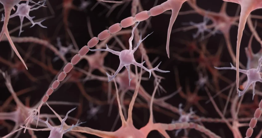
The conventional textbook depiction of a nerve cell’s axon—the long, slender projection responsible for transmitting electrical impulses—as a smooth, unvarying tube may be an oversimplification. New research emerging from Johns Hopkins Medicine, corroborated by subsequent findings in human brain tissue, suggests that many axons exhibit a "pearled" or beaded appearance, characterized by repeating, microscopic swellings. This unexpected architectural feature, distinct from previously understood pathological bulges, appears to play a significant role in modulating the speed and efficiency of neural communication.
The groundbreaking study, published online in Nature Neuroscience on December 2, 2024, utilized advanced imaging techniques to reveal this intricate nanoscale architecture in mouse neurons. This discovery challenges a century of established understanding regarding the morphology of axons, the vital "cables" that connect the vast network of the brain, underpinning functions such as learning, memory, and sensory processing. The implications of these findings extend to our fundamental understanding of neural signaling and could potentially inform future therapeutic strategies for neurological disorders.
Revisiting Axon Anatomy: Beyond the Smooth Tube
For decades, scientific illustrations and models have portrayed axons as relatively uniform, cylindrical structures. While the presence of synaptic varicosities—larger bulges at the terminals of axons that house neurotransmitters for communication with other neurons—has been well-documented, the underlying axon itself was largely assumed to be a consistent conduit. However, the Johns Hopkins team, led by Dr. Shigeki Watanabe, an associate professor of cell biology and neuroscience, observed a different reality in their meticulous examination of mouse neurons.
Using high-pressure freezing electron microscopy, a technique that preserves delicate cellular structures in a near-native state, researchers were able to visualize axons with unprecedented detail. This method, unlike traditional chemical fixation and dehydration, offers a more accurate representation of the axon’s morphology. "To see nanoscale structures with standard electron microscopy, we fix and dehydrate the tissues, but freezing them retains their shape—similar to freezing a grape rather than dehydrating it into a raisin," explained Dr. Watanabe.
Across tens of thousands of images derived from both cultured mouse neurons and tissue from adult and embryonic mice, the researchers repeatedly encountered these minute, repeating swellings, which they termed "non-synaptic varicosities." Crucially, these structures were observed in axons that were not undergoing disease processes and were not associated with synaptic transmission sites. This distinction is paramount: the observed pearling was not indicative of damage but rather suggested an integral component of normal axon architecture, particularly in nonmyelinated axons.
A Trail of Clues: From Worms to Mammalian Brains
Dr. Watanabe’s initial curiosity regarding this pearled morphology was sparked by earlier observations of similar structures in the axons of simpler organisms, such as worms. A discussion with Swiss scientist Dr. Graham Knott, a collaborator on the project, further fueled the investigation. One early hypothesis explored the role of the axon’s internal cytoskeleton, a scaffolding of proteins that provides structural support. Previous research from a Harvard University team in 2012 had highlighted repeating skeletal components within axons.
However, experiments conducted by Jacqueline Griswold, a Johns Hopkins graduate student and the study’s first author, aimed at disrupting this framework yielded a surprising result: the pearling persisted. This finding redirected the research focus towards the physical properties of the axon membrane itself, suggesting that external forces might be shaping the axon’s structure.
To delve deeper into this, Dr. Watanabe and Ms. Griswold collaborated with theoretical biophysicist Dr. Padmini Rangamani, a professor of pharmacology at the University of California San Diego School of Medicine. Together, they employed mathematical modeling to simulate how the surrounding membrane could influence the axon’s beaded appearance.
Unraveling the Mechanics: Membrane Physics in Action
The team’s computational models, which focused on the mechanical properties of the axon’s outer membrane, successfully replicated key features of the observed pearling. This indicated that physical forces inherent to the membrane could be responsible for creating these bulges without necessarily relying on a rigid internal structural element.
Further experimental validation supported this hypothesis. When the sugar concentration in the surrounding solution of the cultured axons was increased, leading to higher osmotic pressure, the swollen regions became smaller. This observation aligned with the models, which predicted that increased membrane tension would reduce the size of the varicosities.
The researchers also manipulated the cholesterol content of the membrane. Cholesterol plays a critical role in membrane fluidity and stiffness. By reducing cholesterol, the membrane became less stiff and more fluid. This alteration had a demonstrable effect on the pearled structure, both in the mathematical models and in the actual mouse neurons. Significantly, this change in membrane fluidity also led to a slower transmission of electrical signals along the axons. Dr. Watanabe elaborated on this, stating, "A wider space in the axons allows ions [chemical particles] to pass through more quickly and avoid traffic jams." However, the relationship between the size of the pearls and signaling speed proved complex; the dimensions of both the swellings and the constrictions between them were critical factors.
Activity-Dependent Structural Changes: A Dynamic Axon
Perhaps one of the most compelling findings was the demonstration that neural activity could dynamically alter axon structure. Following high-frequency electrical stimulation, the pearled regions of the axons exhibited a measurable increase in size—an average lengthening of 8% and a widening of 17%. This structural modification persisted for at least 30 minutes after the stimulation ceased.
Concurrently, the researchers observed a slowing of electrical signaling along these stimulated axons, with the effect lasting for at least an hour. The study also revealed that when cholesterol had been depleted from the membrane prior to stimulation, while some structural changes still occurred, the pearling remained less pronounced, and the significant slowing of electrical signals was not detected. This suggests a direct link between membrane composition, axon morphology, and the speed of neural communication. These findings challenge the long-held notion of axons as static conduits, revealing them as dynamic structures capable of responding to electrical activity and actively regulating signal propagation.
Human Corroboration: Pearled Axons in Human Brain Tissue
The initial findings from the Johns Hopkins study were met with significant scientific interest. The researchers had already planned to investigate whether these pearled axon structures were present in human brain tissue. Subsequent research, published online in Neuron on November 24, 2025, and in its February 4, 2026 issue, provided the crucial human validation.
This related study, co-led by Chelsy R. Eddings and Dr. Watanabe, examined human cortical tissue obtained from individuals undergoing epilepsy surgery. By combining electrical stimulation with rapid freezing techniques, similar to those employed in the mouse studies, the researchers captured transient membrane changes in both mouse and human brain slices. Alongside the pearled axons, they observed evidence of ultrafast endocytosis, a process vital for the rapid retrieval of neuronal membrane after the release of chemical signals at synapses.
While these findings extend the observed phenomenon to human tissue, the researchers emphasize that they do not suggest every axon possesses this precise morphology or that pearling has identical functional consequences across the entire human brain. The human study primarily focused on membrane recycling at synapses, but the presence of pearled axons in this context is highly significant.
Broader Implications and Future Directions
The implications of this discovery are far-reaching. Understanding the precise relationship between axon structure and function is fundamental to comprehending how the brain processes information and adapts. This knowledge could be instrumental in deciphering the mechanisms underlying various neurological disorders, where disruptions in neural signaling are a hallmark.
The importance of this research has been recognized through a significant grant from the National Institute of Mental Health (NIMH). A Multiple Principal Investigator grant awarded to Dr. Watanabe and Dr. Rangamani in 2024 supports further computational modeling of how a neuron’s physical properties and incoming signals influence its axon structure, followed by experimental validation in animal models and cell cultures. This integrated approach promises to deepen our understanding of the intricate interplay between biophysics and neural function.
Furthermore, other research initiatives are contributing to the broader exploration of axon morphology. A study published in PLOS Biology on July 17, 2026, by researchers at the University of Edinburgh, employed automated imaging of living zebrafish to screen a large library of chemical compounds for their effects on axon diameter. They identified several promising compounds that could influence axon width by affecting specific chemical signaling pathways. While not a direct replication of the pearling study, this work provides valuable complementary tools for investigating the complex regulatory mechanisms governing axon size.
For the Johns Hopkins team, a critical next step remains to elucidate the connection between the pearled structures observed in healthy, functioning axons and the more pronounced beading associated with neurodegenerative diseases. Differentiating between normal structural plasticity and pathological alterations could provide crucial diagnostic markers and unlock new avenues for therapeutic intervention. The ongoing investigation into the brain’s microscopic wiring promises to reshape our understanding of neural communication and its susceptibility to disease, offering hope for advancements in treating conditions that affect millions worldwide.

0 Comments