
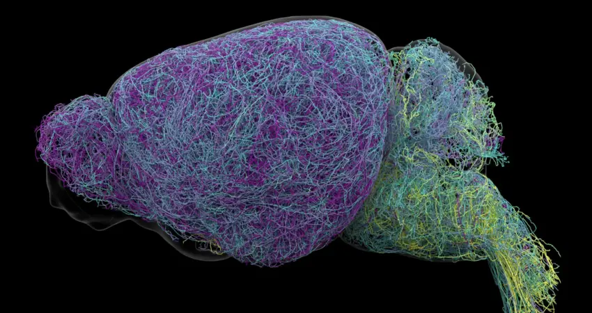
Deep within the brainstem lies a minuscule cluster of nerve cells, the locus coeruleus (LC), whose influence on brain function is disproportionately vast. Its name, derived from Latin for "blue place," hints at its elusive nature. Despite comprising a mere fraction of the brain’s neuronal population, the LC is the sole source of norepinephrine (NE), a critical neurotransmitter orchestrating attention, learning, heart rate regulation, and the body’s dynamic responses to stress and excitement. For decades, the scientific community operated under the assumption that the LC functioned as a broad broadcast system, disseminating a uniform NE signal across the entire brain. However, groundbreaking research published in the prestigious journal Nature has fundamentally reshaped this understanding, unveiling a sophisticated and targeted communication network.
Unraveling the Locus Coeruleus’s Hidden Network
A collaborative effort by researchers at the Allen Institute, employing a sophisticated arsenal of techniques including advanced brain imaging, precise neural activity recordings, cutting-edge genetic methodologies, and meticulously designed behavioral experiments in mice, has revealed that the LC communicates through multiple distinct pathways. This paradigm-shifting discovery indicates that the LC does not merely issue a general alert but rather delivers specific signals to targeted destinations, with different neuronal populations exhibiting differential responses to varied experiences.
A Sophisticated Postal Network, Not a Foghorn
Through meticulous tracing of LC neuron connections, scientists have mapped out how distinct groups of these cells engage with specific regions of the nervous system. These identified pathways precisely dictate where NE is released, thereby establishing a far more organized and nuanced communication infrastructure than previously conceived.
The LC exhibits a clear topographical organization. Neurons situated in its upper, dorsal portion predominantly establish connections with the cerebral cortex and the forebrain. Conversely, cells residing in the lower, ventral region of the LC primarily project their signals to the brainstem and the spinal cord. This anatomical segregation empowers different segments of the LC to exert influence over distinct neural functions.
"The findings suggest the brain’s norepinephrine system is far more like a targeted postal network than a foghorn," stated Karel Svoboda, director of Neural Dynamics at the Allen Institute and a co-author of the study. This analogy effectively captures the transition from a generalized broadcast model to one characterized by precision delivery and specific targeting.
Implications for Neurological and Mental Health Disorders
The profound implications of this discovery extend to the potential transformation of how certain neurological and mental health conditions are understood and treated. Norepinephrine’s pivotal role in regulating attention and mood makes it a primary target for pharmacological interventions in conditions such as depression, anxiety, and Attention-Deficit/Hyperactivity Disorder (ADHD).
Furthermore, LC neurons are among the earliest to exhibit degeneration in Alzheimer’s disease. This observation strongly suggests that the new insights into the LC’s organizational structure could prove invaluable in unraveling the complexities of and developing effective treatments for neurodegenerative disorders.
Current therapeutic strategies that modulate norepinephrine activity often result in widespread effects across multiple brain circuits. While these treatments may offer benefits in one neural region, they can concurrently induce undesirable side effects in others. The detailed mapping of the LC’s specialized pathways provided by this research offers the tantalizing prospect of developing therapies that can selectively target the specific neural circuits implicated in particular symptoms, thereby enhancing efficacy and minimizing adverse reactions.
The research that illuminated these critical brain pathways was generously supported by the National Institutes of Health’s Brain Research Through Advancing Innovative Neurotechnologies® Initiative, widely recognized as The BRAIN Initiative®.
Decoding Neuronal Roles in Learning and Engagement
Beyond mapping the structural organization of the LC, the researchers also identified a crucial functional distinction between different subsets of LC neurons.
Neurons within the dorsal LC, those that project to the cerebral cortex, demonstrated particularly heightened activity during learning processes. Specifically, when mice engaged in decision-making tasks and subsequently modified their choices following an unfavorable outcome, these dorsal LC neurons exhibited a significant increase in their firing rates. This suggests a direct involvement in adaptive learning and behavioral adjustment.
In contrast, neurons in the ventral LC displayed a different activity pattern. Their activity surged just prior to mice disregarding signals that indicated the potential availability of a reward. This observation points towards a regulatory function in controlling an organism’s propensity to respond to opportunities presented by its environment, playing a role in engagement or disengagement with external stimuli.
"What emerged was a clear map: neurons in the dorsal LC that send signals upward to the cortex are involved in learning, whereas neurons in the ventral region projecting downward to the brainstem and spinal cord govern whether animals engage with their environment at all," explained Svoboda. He further elaborated, "We also showed that these anatomical differences are mirrored by distinct gene expression patterns." This finding underscores that the divergence between these neuronal groups is not merely anatomical but is also reflected in their unique molecular profiles, reflecting their specialized roles.
A Remarkable Discovery: The Record-Breaking Neuron
The comprehensive nature of this study involved an extensive examination of the mouse brain. Scientists employed whole-brain imaging techniques to meticulously analyze nearly 35,000 neurons. Concurrently, they analyzed the genetic profiles of almost 400,000 cells and undertook the challenging task of reconstructing the complete intricate structures of several exceptionally complex nerve cells.
During this monumental undertaking, an additional, astonishing discovery was made: LC neurons possess extraordinarily elongated axons. Axons are the critical projections that transmit signals from one nerve cell to another, thereby facilitating the transmission of information throughout the nervous system.
The research team found that the average length of LC axons measured approximately 35 centimeters. In a truly exceptional case, one reconstructed axon extended to an astonishing 70.32 centimeters, establishing it as one of the longest single neurons ever measured in a mouse.
"This neuron, like many others that we studied, supplies NE to a very large volume of the cerebral cortex. For the brain, this is highly unusual. Most neurons are more specific in their targets," commented Jeremiah Cohen, a scientist at the Allen Institute and co-author of the study. He further clarified the selective nature of this extensive reach: "But this neuron doesn’t release NE everywhere. It ignores the cerebellum, brainstem, and spinal cord." This observation further reinforces the central finding that, despite their remarkable physical extent, LC neurons do not indiscriminately broadcast norepinephrine. Instead, their connectivity remains remarkably selective, underscoring the sophisticated organization of the LC’s signaling pathways.
Forging a New Understanding of Brain Learning Mechanisms
The research also unveiled intriguing parallels between the norepinephrine signaling system and the brain’s dopamine signaling network. Dopamine, another pivotal chemical messenger, transmits signals to the basal ganglia, a brain region integral to the formation of habits and reward-based learning. Scientists hypothesize that dopamine and norepinephrine may collaborate synergistically to support learning processes, potentially enabling the brain to acquire and consolidate a diverse range of complex and abstract concepts simultaneously.
By meticulously delineating the intricate connectivity, specialized functions, and unusual physical architecture of neurons within the locus coeruleus, this research provides an unprecedentedly detailed depiction of how the mammalian brain communicates and learns. While the findings originate from experiments conducted on mice, their potential to enhance our understanding of human brain function and to guide the development of more precise therapeutic interventions targeting specific neural circuits, rather than broad signaling systems, is immense. This work marks a significant leap forward in our quest to unravel the brain’s most fundamental processes.

0 Comments