
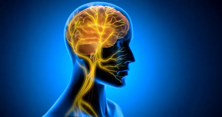
The perplexing question of why some skills are acquired with remarkable ease while others resist even dedicated practice may soon have a clearer answer. Beyond innate talent and sheer effort, a crucial factor in the persistence of new knowledge might lie within the brain’s internal readiness to transform fleeting experiences into enduring memories. New research from Tohoku University, published on August 25, 2026, in the journal iScience, illuminates a previously underestimated role for the body’s internal signaling system, specifically the vagus nerve, in solidifying motor learning in mammals. This groundbreaking study, conducted on mice, suggests that stimulating this vital communication pathway after training can significantly bolster the brain’s capacity for long-term skill retention, pointing to a potent, yet largely overlooked, brain-body dialogue.
The Vagus Nerve: A Bridge Between Internal States and Cognitive Function
The vagus nerve, the longest cranial nerve, acts as a sophisticated bidirectional communication channel, constantly relaying information from the body’s internal organs to the brain, and vice versa. This intricate network plays a pivotal role in regulating numerous bodily functions, including heart rate, digestion, and even mood. In recent years, the scientific community has begun to explore its potential in neuromodulation, with vagus nerve stimulation (VNS) already an established clinical therapy for conditions such as epilepsy and depression. However, the Tohoku University study proposes a novel mechanism, moving beyond the traditional understanding of VNS as a broad modulator of neurotransmitter systems. Researchers now hypothesize that VNS might exert its learning-enhancing effects by influencing rhythmic changes within the brain’s vascular network, a subtle but significant shift in our comprehension of its impact on cognitive processes.
A Novel Approach to Investigating Learning Enhancement
To explore this hypothesis, the research team developed a specialized cuff electrode designed for precise attachment to the left cervical vagus nerve of laboratory mice. The study focused on the horizontal optokinetic response (HOKR) learning task. This cerebellum-dependent motor skill involves teaching mice to track moving visual stripes with their eyes, mimicking the natural compensatory eye movements humans make when observing a moving landscape. The HOKR task was chosen for its well-established link to cerebellar function, a brain region critical for motor learning and memory consolidation.
The experimental design was particularly insightful. Vagus nerve stimulation was not applied during the learning sessions themselves, but rather in the critical period following each training block. This strategic timing was key, as the researchers hypothesized that the benefits would manifest not as immediate performance boosts during practice, but as enhanced retention and consolidation of the learned skill over time.
Post-Training Stimulation Unlocks Durable Memory
The results of the study provided compelling evidence for this hypothesis. Mice that received vagus nerve stimulation after their training sessions demonstrated significantly stronger long-term motor learning compared to control groups. This pattern strongly suggests that VNS, when applied in the post-practice window, engages mechanisms responsible for memory consolidation – the process by which newly acquired information is stabilized and transformed into enduring memories.
Professor Ko Matsui, a key figure in the research, emphasized this critical aspect of the findings: "The key point is that VNS was delivered only after training," he stated. "Our findings suggest that VNS may open a hidden window of opportunity for enhanced learning by making the brain environment more receptive to long-lasting change." This implies that the body’s internal signals, amplified by VNS, can prime the brain to better encode and retain newly acquired motor skills.
Unveiling the Vascular Rhythms of Learning
The research team then delved deeper, seeking to identify the specific physiological changes within the brain that correlated with the observed improvements in learning. They employed advanced techniques, including fiber photometry, to monitor blood volume activity in the vicinity of the cerebellar flocculus, a region known to be crucial for HOKR learning.
Their investigations revealed a fascinating two-phase vascular response following a single instance of VNS. Initially, local blood volume experienced a brief decrease, followed by a delayed increase. When VNS was administered repeatedly, this vascular response evolved into distinct rhythmic oscillations in blood volume. This finding was particularly significant because these vascular rhythms appeared to be directly linked to the learning outcomes. The study observed a strong correlation: mice exhibiting more pronounced blood volume oscillations generally demonstrated superior learning by the fifth day of the experiment. This suggests that the dynamic changes in the brain’s vascular environment, orchestrated by VNS, are a critical component in facilitating the lasting effects of the stimulation on learning.
"Our brains may be more strongly influenced by the body than we imagine," commented lead author Junyu Chen. "By tuning the brain’s metabolic environment, including rhythmic vascular movements, we may eventually unlock capacities that would otherwise remain latent." This statement highlights the profound implications of the research, suggesting that by understanding and manipulating the intricate interplay between the body and brain, we may be able to tap into previously unrecognized potential for cognitive enhancement.
Broader Implications and Future Directions
The implications of this research extend far beyond the laboratory setting. The discovery that stimulating the vagus nerve after practice can enhance long-term motor learning opens up exciting avenues for developing novel therapeutic and educational interventions.
Supporting Data and Context:
While specific quantitative data on learning improvement percentages were not detailed in the initial release, the observed correlation between the magnitude of vascular oscillations and learning outcomes by Day 5 provides a robust indication of the VNS effect. Further studies will likely quantify the degree of improvement in skill retention, comparing it to baseline learning rates and other known learning enhancement techniques. Historically, research into learning consolidation has focused heavily on neural plasticity mechanisms, such as long-term potentiation (LTP). This new research adds a significant physiological dimension by highlighting the role of vascular dynamics, potentially offering a complementary or even synergistic pathway for enhancing these neural processes.
Timeline and Chronology:
The research leading to this publication likely spanned several years, involving experimental design, data acquisition, analysis, and peer review. The specific timeline within the study involved:
- Initial Setup: Development of the VNS cuff electrode and establishment of the HOKR learning paradigm in mice.
- VNS Application: Application of VNS after daily training sessions over a defined period.
- Performance Monitoring: Assessment of HOKR performance during training and in subsequent retention tests.
- Vascular Monitoring: Measurement of blood volume changes in the cerebellum using fiber photometry during and after VNS.
- Data Analysis: Correlation of VNS application, vascular rhythms, and learning performance.
- Publication: Dissemination of findings in iScience on August 25, 2026.
Potential for Enhanced Rehabilitation and Training:
The findings could revolutionize approaches to physical rehabilitation for individuals recovering from stroke, traumatic brain injury, or other neurological conditions that impair motor function. By integrating VNS protocols into rehabilitation regimens, therapists might accelerate motor skill recovery and improve the long-term functional outcomes for patients. Similarly, in athletic training and skill acquisition for complex tasks, targeted VNS could potentially optimize the learning curve and enhance the durability of learned skills.
Understanding Brain-Body Communication:
This study underscores the profound and often subtle ways our bodies communicate with our brains, influencing our cognitive abilities. It challenges a view of the brain as an isolated processing unit and emphasizes its integration within a larger physiological system. The rhythmic vascular changes observed suggest that the brain’s metabolic environment, influenced by bodily signals, is a critical substrate for learning and memory.
Future Research Directions:
The researchers are keen to expand upon these findings. Future investigations will likely focus on:
- Optimizing VNS Protocols: Determining the ideal frequency, intensity, and duration of VNS for different types of learning and in various biological contexts.
- Precise Mechanisms: Elucidating the exact molecular and cellular pathways through which vagal nerve stimulation influences vascular dynamics and neuronal plasticity.
- Translational Studies: Exploring the applicability of these findings in human subjects, potentially using non-invasive VNS techniques.
- Exploring Other Sensory Inputs: Investigating whether other forms of bodily feedback can similarly influence learning and memory consolidation.
By continuing to explore this vital two-way communication pathway between the brain and body, scientists aim to unravel the fundamental mechanisms of lasting learning and, ultimately, to develop innovative strategies for enhancing human cognitive capabilities throughout the lifespan. This research marks a significant step forward in understanding the holistic nature of learning and the untapped potential of the brain-body connection.

0 Comments