
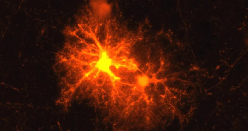
When laboratory mice sustain brain damage, such as that induced by an injection, Jan Deussing, a seasoned neurobiologist and research group leader at the Max Planck Institute of Psychiatry, has consistently observed a specific cellular response. A distinct population of cells congregates and becomes highly active in the vicinity of the injured area. Despite repeated observations of this phenomenon, the precise identity of these crucial cells remained an enigma for Deussing. This persistent mystery presented an ideal challenge for a budding scientist. Clemens Ries, a master’s student nearing the completion of his biology degree and undertaking an internship at the institute, embraced this intriguing research question.
The Unveiling of the Brain’s Progenitor Cells
Ries embarked on a systematic investigation, employing a mouse model and meticulously testing a comprehensive array of cell markers representing all known cell types within the brain. His rigorous screening process yielded a singular, decisive result: a marker specifically associated with oligodendrocyte progenitor cells (OPCs) consistently elicited a response around the damaged brain tissue.
These OPCs are a fundamental component of the central nervous system’s cellular landscape. They serve as precursor cells, possessing the remarkable capacity to mature into oligodendrocytes. These mature cells are the architects of the myelin sheath, a vital insulating layer that encases the axons of nerve cells. Axons are the long, slender extensions of neurons responsible for transmitting electrical and chemical signals, enabling communication throughout the intricate neural network. Analogous to the insulation surrounding an electrical cable, myelin plays a critical role in facilitating efficient and rapid information transmission along axons. Furthermore, myelin provides essential metabolic support to these neuronal extensions, a function indispensable for maintaining overall brain health and optimal neural function.
The integrity of the myelin sheath is paramount. Damage to this protective coating can precipitate a cascade of severe neurological consequences. In debilitating autoimmune diseases like multiple sclerosis (MS), the immune system erroneously attacks and degrades the myelin sheath, leading to widespread neurological dysfunction. Physical trauma to the brain can also inflict significant damage on myelin, and in severe instances, this injury can result in the irreversible death of entire neurons. Consequently, the brain’s inherent capacity to repair and regenerate myelin around compromised axons represents a critical aspect of its restorative processes following injury.
A Surprising Stress Hormone Emerges in the Wake of Injury
Initially, Ries’s exploration of these newly identified cells was confined to his master’s thesis. However, the profound implications and the sheer scientific intrigue of the findings compelled him to delve deeper. "The topic remained so exciting that it became my doctoral thesis," Ries stated, underscoring the captivating nature of his research journey.
His subsequent doctoral research revealed a dramatic proliferation of these progenitor cells at the periphery of brain lesions. Crucially, a significant proportion of these OPCs then embarked on a maturation pathway, transforming into fully functional oligodendrocytes capable of synthesizing new myelin. This discovery offered a glimpse into the cellular mechanisms underlying brain repair.
However, Ries and Deussing’s investigation uncovered an even more unexpected revelation. In close proximity to the injured tissue, approximately one-third of the OPCs exhibited the activation of corticotropin-releasing hormone (CRH). CRH is a neuropeptide primarily recognized for its central role in orchestrating the body’s physiological and behavioral responses to stress. Prior to this research, it was not known that OPCs possessed the capability to produce such neuropeptides. The groundbreaking findings of this study were subsequently published in the esteemed scientific journal Cell Reports, marking a significant advancement in the field of neurobiology.
The onset of the CRH response proved to be remarkably swift. Detectable levels of CRH production could be observed within a mere few hours following an injury. However, this surge in CRH expression was transient, subsiding after approximately three days. This short, intense burst of activity strongly suggests that CRH plays a pivotal role during the very earliest stages of the brain’s healing response, potentially priming the cellular environment for subsequent repair.
CRH: A Key Regulator of Myelin Repair Timing
Further investigation revealed that one of the two known receptors for CRH, specifically CRH receptor 1 (CRHR1), plays a central role in this intricate process. CRHR1 is expressed on a distinct subpopulation of OPCs, enabling these cells to respond to the CRH released by their neighboring counterparts.
When CRHR1 is absent or non-functional, OPCs demonstrate an accelerated rate of proliferation in the aftermath of an injury. Paradoxically, this initial surge in cell numbers does not translate into more effective myelin repair. Instead, the research indicated that in the absence of CRHR1, fewer mature oligodendrocytes are ultimately produced and sustained, suggesting a critical role for CRH signaling in optimizing the repair process.
These findings strongly indicate that CRH acts as a crucial regulator, meticulously controlling the timing of OPC maturation. This precise temporal coordination appears to be essential for ensuring the generation of a sufficient number of mature oligodendrocytes, which are then tasked with the vital job of restoring the damaged myelin sheath. Without this regulated timing, the repair process may be inefficient, leading to incomplete or suboptimal myelination.
The Developmental Echo: CRH and Brain Maturation
The significance of OPCs extends far beyond their role in injury repair. They are also instrumental in the fundamental process of myelin formation during normal brain development, a period that commences in utero and continues well into young adulthood. A substantial portion of this critical myelination process occurs postnatally, laying the groundwork for cognitive function and behavioral development.
Given that CRHR1 is present on OPCs even in the absence of any injury, Ries and Deussing hypothesized that this receptor might also influence myelination during the brain’s developmental trajectory. Collaborating with other research teams, they employed a variety of sophisticated experimental methods to examine myelin formation in additional mouse models.
Their research uncovered compelling evidence that mice lacking functional CRHR1 exhibited an increased number of OPCs during the early stages of brain development. These developmental alterations were not transient; they persisted with age, exerting lasting effects on the overall structure of the brain. In adult brains of these mice, researchers observed measurable changes in myelination patterns, notably characterized by thicker myelin sheaths, particularly around thinner axons. These results strongly suggest that CRH receptor 1 on OPCs is not only vital for myelin repair following injury but also plays a fundamental role in regulating the intricate process of myelin development itself.
The Source of CRH During Development: A Neuronal Connection
While injury triggers OPCs to produce and release CRH, the origin of this stress hormone during normal brain development presents a different set of questions. The scientists have put forth a compelling hypothesis: developing neurons themselves may be the primary source of CRH during this critical period. Their proposed model suggests that as neurons mature and establish connections, they release CRH, which in turn influences both the proliferation of OPCs and their subsequent differentiation into myelin-producing oligodendrocytes. This proposed mechanism offers a novel perspective on how neuronal activity and cellular differentiation are intricately linked during brain maturation.
Implications for Mental Health: Stress, Depression, and the Developing Brain
The established role of neurons in releasing CRH, particularly under stressful conditions, coupled with the known association between early-life stress and an increased risk of psychiatric disorders, lends significant weight to the broader implications of these findings. The newly elucidated CRH system operating within OPCs raises the possibility of a more profound connection between stress signaling, myelin integrity, and mental health than previously understood.
"Our current findings suggest that in stress-associated psychiatric disorders such as depression, the CRH system in OPCs may play a greater role than previously known," Deussing speculates, highlighting the potential paradigm shift these discoveries could represent. If future research definitively confirms and expands upon this connection, a deeper understanding of how CRH signaling impacts OPCs, myelin formation, and overall brain development could pave the way for entirely novel therapeutic strategies. These could potentially target the CRH-OPC axis to address the underlying neurobiological deficits associated with various psychiatric conditions, offering hope for improved treatments and interventions. The complex interplay between stress, cellular repair mechanisms, and brain development is a frontier of research with immense potential to unlock new avenues for understanding and treating debilitating neurological and psychiatric disorders.

0 Comments