
Before birth, the human brain undergoes an extraordinary and intricate construction, orchestrated by a cascade of cellular decisions. At the heart of this monumental process lie radial glia, a specialized class of neural stem cells instrumental in forging the very architecture that distinguishes the human brain. These remarkable cells are the primary architects, generating the vast numbers of neurons and essential support cells that constitute the cerebral cortex—the brain’s command center for cognition, memory, and language. Radial glia are also strongly implicated in the dramatic expansion of the human cortex, a key differentiator when compared to the brains of other species. While most of these critical cells typically vanish before birth, a perplexing phenomenon sees similar cells reappear in brain cancers, a reappearance whose underlying causes remain an active area of scientific investigation.
"Radial glia are the coolest cells that have ever existed," stated Aparna Bhaduri, an assistant professor of biological chemistry at the David Geffen School of Medicine at UCLA. "They’re really key to making us human. But they’re also at the center of many neurodevelopmental and neuropsychiatric disorders, as well as cancer—so understanding how they make their decisions is one way to start understanding how those conditions arise."
Two groundbreaking new studies, published concurrently in the prestigious journals Cell and Science, are now illuminating the intricate decision-making processes of radial glia during development. The research, spearheaded by Bhaduri and her colleagues, reveals that these pivotal cells are exquisitely sensitive to two distinct streams of information: the way they process nutrients and direct physical signals originating from other developing brain regions. Collectively, these findings offer unprecedented insights into the mechanisms by which the human cortex achieves its remarkable diversity of cell types, a complexity that underpins human intelligence.
Metabolic Pathways Dictate Neural Stem Cell Fate
The study published in Cell represents a significant leap forward in understanding the metabolic landscape of the developing human cortex. This ambitious project, a collaborative effort between Bhaduri’s lab and the laboratory of Heather Christofk, was co-led by Jessenya Mil and Jose Soto.
To construct this comprehensive atlas, the research team meticulously analyzed both donated human brain tissue and sophisticated brain organoids meticulously grown from stem cells. Their findings yielded a surprising conclusion: metabolism is not merely a passive backdrop supporting brain development. Instead, it actively intervenes, profoundly influencing the types of cells that are ultimately produced.
A key discovery was the crucial dependence of radial glia on the pentose phosphate pathway. This metabolic pathway is a vital cellular engine, utilizing glucose to generate essential building blocks required by cells that are undergoing rapid proliferation.
When the researchers experimentally reduced the availability of glucose or disrupted the functioning of this pathway, a tangible shift occurred in the cells’ output. The radial glia began to generate a higher proportion of inhibitory neurons and other cell types that typically emerge later in the developmental timeline. This observation underscores the dynamic and directive role of cellular metabolism.
"What was surprising is that metabolism isn’t just a passive thing that happens in the background," commented Bhaduri, who is also affiliated with the UCLA Broad Stem Cell Research Center and the UCLA Health Jonsson Comprehensive Cancer Center. "It can really control how stem cells make decisions."
These groundbreaking results have profound implications for understanding how external factors influence brain development. They open new avenues for investigating the impact of maternal nutrition, metabolic disorders, and other environmental influences on the developing human brain. The metabolic atlas itself stands as one of the most detailed resources available to date for researchers delving into the complexities of metabolism during human brain development.
Early Thalamic Signals: A Physical Blueprint for Cortical Development
The second study, featured in Science and led by first author Claudia Nguyen, explored an entirely different category of developmental cues: signals emanating from the thalamus. This structure, nestled deep within the brain, acts as a critical relay station, transmitting sensory and motor information throughout the nervous system.
For years, neuroscientists have known that thalamic neurons extend long axonal projections towards the developing cortex. These thread-like fibers are destined to form intricate connections with specific cortical neurons. However, anatomical studies have revealed a striking human-specific characteristic: these thalamic projections reach the cortex significantly earlier than the final synaptic connections are established. This temporal discrepancy posed a compelling question: what is the purpose of these premature arrivals?
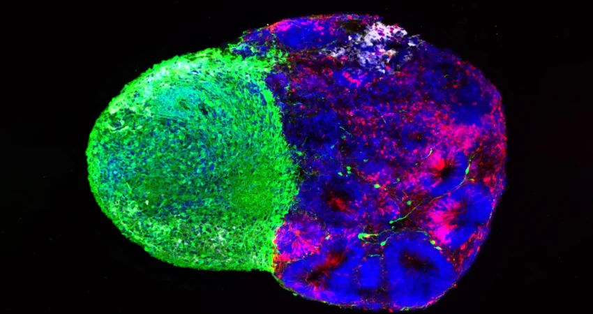
Employing advanced human stem cell-derived brain "assembloids"—complex in vitro models that recapitulate key aspects of brain development—the UCLA researchers uncovered a crucial part of the answer. They demonstrated that these thalamic projections physically interact with radial glia during the embryonic stages of brain formation.
This direct physical contact profoundly altered the behavior of the neural stem cells. It stimulated them to produce a greater number of excitatory neurons, the primary workhorses responsible for transmitting signals within the cortex. This effect was particularly pronounced for upper-layer neurons, a class of neurons that are notably expanded in the human brain, contributing to its advanced cognitive capabilities.
"We already knew that these projections influence how the cortex develops," Bhaduri explained. "What we specifically found is that this influence comes through an actual physical connection between the projections and the radial glia—a point of contact that just hasn’t been identified before, and one that very likely does not exist in rodents." This suggests a specialized mechanism for human cortical development.
The NRXN1 Gene: A Link to Autism and Developmental Precision
Further investigation by the research team revealed a critical molecular player in this thalamic-cortical interaction: the NRXN1 gene. This gene is already well-established for its role in facilitating the formation of neural connections. Importantly, mutations in NRXN1 have been previously linked to autism spectrum disorder (ASD), highlighting its significance in neurodevelopment.
To explore the gene’s function in this context, the researchers engineered assembloids using patient-derived cells carrying an NRXN1 mutation. In these experimental models, the altered thalamic signals exhibited distinct behaviors compared to signals generated by unaffected cells.
These molecular changes led to a disruption in the delicate balance between the population of stem cells and the neurons they were generating. This finding offers a promising avenue for researchers to investigate how subtle disturbances early in brain development, potentially mediated by genetic factors like NRXN1 mutations, could profoundly influence the intricate architecture of the developing cortex and contribute to neurodevelopmental disorders.
A Symphony of Signals: The Developing Brain in Constant Dialogue
While these two studies focused on markedly different mechanisms—one on the internal metabolic environment of the cells and the other on external physical and molecular signals from another brain region—they converge on a singular, overarching principle: radial glia do not make their crucial developmental decisions in isolation. Their fate and function are continuously shaped by a dynamic interplay of signals from their immediate environment.
Furthermore, these studies underscore the transformative power of organoid technology in advancing the study of human brain development. Just a decade ago, direct investigation into the behavior of uniquely human neural stem cells was severely limited by the lack of suitable experimental models.
Today, the development of brain organoids and related sophisticated in vitro systems allows scientists to faithfully recreate crucial features of human brain development in the laboratory. These powerful models also enable researchers to address complex questions that are intractable using traditional animal models alone, bridging a critical gap in our understanding.
Bhaduri expressed hope that these findings will inspire a paradigm shift in how scientists perceive the fundamental processes of brain development. She advocates for viewing metabolism and physical cellular interactions not as passive background processes but as active, directive forces that sculpt neural circuits.
"Ultimately, these studies give us a glimpse under the hood of how these cells make decisions," she concluded. "Understanding those decisions is a first step toward understanding normal brain development, disease vulnerability and, potentially, how similar stem-cell programs operate in brain cancer." The implications extend beyond understanding development, offering potential insights into the origins of neurological disorders and the cellular mechanisms underlying brain tumors.
This pioneering research was made possible through substantial support from a consortium of leading scientific organizations, including the National Institutes of Health, the National Science Foundation, the Brain & Behavior Research Foundation, the Alfred P. Sloan Foundation, the Rose Hills Foundation, the Esther A. & Joseph Klingenstein Fund, the Simons Foundation, the Chan Zuckerberg Initiative, the NIH BRAIN Initiative Cell Atlas Network, the International Foundation for Ethical Research, and key UCLA centers: the Broad Stem Cell Research Center (through its Stem Cell Research Training Program) and the UCLA Health Jonsson Comprehensive Cancer Center and UCLA Broad Stem Cell Research Center Ablon Scholars Program.

0 Comments