
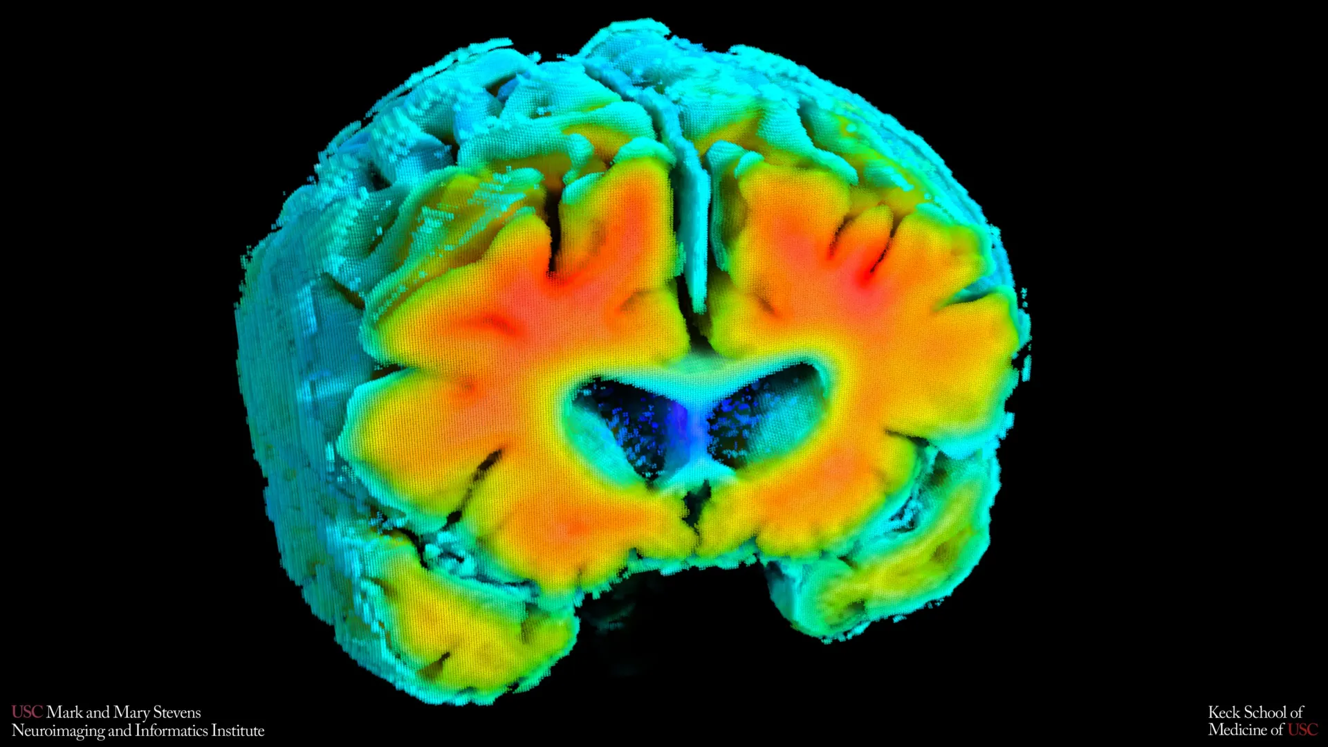
Small shifts in how blood moves through the brain and how brain cells receive oxygen may be closely connected to the risk of Alzheimer’s disease. This is the conclusion of new research from the Mark and Mary Stevens Neuroimaging and Informatics Institute (Stevens INI) at the Keck School of Medicine of USC. The study, published in Alzheimer’s and Dementia: The Journal of the Alzheimer’s Association, examined older adults both with and without cognitive impairment. Researchers found that simple, noninvasive measures of brain blood flow and oxygen levels were linked to well-known signs of Alzheimer’s, including amyloid plaque buildup and shrinkage of the hippocampus, the part of the brain that plays a central role in memory. The results suggest that the health of the brain’s blood vessels may influence the disease process early on and could help flag people at risk before noticeable symptoms develop.
"Amyloid and tau are often considered the primary players in Alzheimer’s disease, but blood flow and oxygen delivery are also critical," said Amaryllis A. Tsiknia, lead author of the study and USC PhD candidate. "Our results show that when the brain’s vascular system functions more like it does in healthy aging, we also see brain features that are linked to better cognitive health."
This groundbreaking research offers a potential paradigm shift in how Alzheimer’s disease is understood and detected. For decades, the focus has largely been on the accumulation of abnormal protein deposits – amyloid plaques and tau tangles – as the primary drivers of neurodegeneration. While their role remains undeniable, this new study emphasizes the integral contribution of the brain’s circulatory system, suggesting that vascular health may be a foundational element in the Alzheimer’s continuum, potentially preceding the more widely recognized pathological hallmarks.
Noninvasive Tools Illuminate Brain Circulation Dynamics
The research team employed two innovative, noninvasive techniques to meticulously assess the intricate mechanisms of brain circulation. These methods were selected for their accessibility and ease of use, allowing for objective measurements without requiring invasive procedures or extensive patient discomfort.
One of the key tools utilized was Transcranial Doppler (TCD) ultrasound. This technique employs sound waves to measure the speed of blood flow through the major arteries supplying the brain. By analyzing the velocity of blood as it travels through these crucial vessels, researchers can gain insights into the overall patency and efficiency of the brain’s arterial network. Blood flow velocity is a sensitive indicator of vascular health; reduced flow can signal narrowing or stiffening of arteries, a common consequence of aging and various cardiovascular conditions that can also impact the brain.
Complementing the TCD ultrasound, the researchers also utilized Near-Infrared Spectroscopy (NIRS). NIRS is a non-invasive optical imaging technique that assesses tissue oxygenation by measuring the absorption and scattering of near-infrared light. In the context of this study, NIRS was used to evaluate how effectively oxygenated blood reaches the superficial layers of the cerebral cortex. The brain is an incredibly metabolically active organ, demanding a constant and robust supply of oxygen to function. Impaired oxygen delivery can lead to cellular stress and dysfunction, contributing to neurodegenerative processes.
Advanced Modeling Enhances Vascular Assessment
Beyond simply collecting raw data from these instruments, the USC team applied sophisticated advanced mathematical modeling to integrate the readings. This computational approach allowed them to synthesize the TCD and NIRS data into comprehensive indicators of cerebrovascular function. These combined indicators are designed to reflect the brain’s dynamic capacity to regulate blood flow and oxygen delivery. Specifically, they measure how effectively the brain can adapt its circulatory response to physiological fluctuations, such as normal variations in blood pressure and levels of carbon dioxide in the blood.
A healthy cerebrovascular system is characterized by its ability to maintain a stable internal environment for brain cells despite external changes. For instance, during periods of increased metabolic demand or when blood pressure drops slightly, the brain’s vessels can constrict or dilate to ensure consistent blood flow and oxygenation. Conversely, a compromised vascular system may exhibit a diminished ability to make these necessary adjustments, leaving brain tissue vulnerable to periods of insufficient oxygenation or excessive pressure. The mathematical models developed by the researchers aim to quantify this adaptive capacity, providing a more nuanced understanding of vascular health than single-point measurements alone.
Direct Correlation: Vascular Health, Amyloid, and Memory Preservation
The study’s findings revealed a striking correlation between enhanced cerebrovascular function and reduced markers of Alzheimer’s disease pathology. Participants whose vascular indicators more closely mirrored those observed in cognitively healthy individuals exhibited lower levels of amyloid plaque buildup in their brains. Amyloid plaques are one of the hallmark pathologies of Alzheimer’s disease, and their presence is strongly associated with cognitive decline.
Furthermore, these individuals with robust vascular function also tended to have a larger hippocampal volume. The hippocampus, a critical brain structure within the medial temporal lobe, is essential for forming new memories and is one of the earliest regions to be affected by Alzheimer’s disease, leading to the memory loss characteristic of the condition. Shrinkage, or atrophy, of the hippocampus is a well-established indicator of neurodegeneration and Alzheimer’s progression.
"These vascular measures are capturing something meaningful about brain health," stated Meredith N. Braskie, PhD, senior author of the study and assistant professor of neurology at the Keck School of Medicine. "They appear to align with what we see on MRI and PET scans that are commonly used to study Alzheimer’s disease, providing important information about how vascular health and standard brain measures of Alzheimer’s disease risk may be related."
The alignment with established imaging modalities like MRI (Magnetic Resonance Imaging) and PET (Positron Emission Tomography) scans is particularly significant. MRI is used to visualize brain structure, including hippocampal volume, while PET scans can detect amyloid and tau pathology. The fact that these simpler, noninvasive vascular measures correlate with findings from these more complex and costly imaging techniques suggests they offer complementary and potentially early indicators of disease risk.
A Continuum of Decline: Mild Cognitive Impairment and Vascular Function
The research also extended to individuals diagnosed with mild cognitive impairment (MCI) or dementia. MCI is a condition characterized by a noticeable decline in cognitive abilities, such as memory or thinking skills, that is more significant than expected for normal aging but not severe enough to interfere with daily life. It is often considered a prodromal stage for Alzheimer’s disease.
The study observed that individuals with MCI or dementia consistently demonstrated weaker cerebrovascular function when compared to participants who were cognitively normal. This finding strengthens the hypothesis that declining blood vessel health in the brain is not an isolated issue but rather an integral component of the broader Alzheimer’s disease continuum. It suggests that vascular compromise may be an early contributing factor that sets the stage for, or exacerbates, the neurodegenerative processes that define Alzheimer’s.
"These findings add to growing evidence that Alzheimer’s involves meaningful vascular contributions in addition to classic neurodegenerative changes," remarked Arthur W. Toga, PhD, director of the Stevens INI. "Understanding how blood flow and oxygen regulation interact with amyloid and brain structure opens new doors for early detection and potentially prevention."
The implications of this observation are profound. If vascular dysfunction is an early and consistent feature of the Alzheimer’s continuum, then interventions aimed at improving vascular health could potentially serve as a strategy to slow or even prevent the progression of cognitive decline. This perspective broadens the therapeutic landscape beyond solely targeting amyloid and tau.
The Promise of Accessible and Broad-Scale Screening
One of the most compelling aspects of this research lies in the potential for its noninvasive measurement techniques to revolutionize Alzheimer’s screening. Compared to the sophisticated and often expensive imaging modalities like MRI and PET scans, TCD ultrasound and NIRS offer significant advantages in terms of cost-effectiveness, accessibility, and patient comfort.
Cost and Accessibility: MRI and PET scanners represent substantial capital investments for healthcare institutions. Their widespread availability can be limited, particularly in underserved areas. The equipment required for TCD and NIRS is considerably less expensive, making these technologies more feasible for deployment in a wider range of clinical settings, including primary care offices and community health centers.
Patient Comfort and Safety: MRI scans require patients to lie still in a confined space for extended periods, which can be challenging for some individuals, especially those with claustrophobia. PET scans typically involve the injection of a radioactive tracer, which, while generally safe, carries inherent risks and requires specialized handling and disposal. TCD ultrasound and NIRS are entirely noninvasive, painless, and do not involve radiation exposure. They can be performed while a patient is resting quietly, making them ideal for a broader demographic, including the elderly and individuals with mobility issues or anxieties about medical procedures.
Reduced Burden on Patients: The simplicity of these methods reduces the burden on patients, eliminating the need for complex preparations or demanding tasks. This ease of use could make them particularly valuable for large-scale screening initiatives, allowing for the assessment of a greater number of individuals at risk for Alzheimer’s disease. For individuals who are unable to undergo more intensive brain imaging due to medical contraindications or logistical challenges, these noninvasive vascular assessments could provide a crucial pathway to understanding their cognitive health risks.
Future Directions and Long-Term Implications
While the current findings are highly promising, the researchers are mindful of the study’s limitations and the need for further investigation. The study captured a single snapshot in time, meaning it establishes associations but does not definitively prove cause and effect. It is crucial to determine whether observed changes in vascular function precede or merely accompany the development of Alzheimer’s pathology and cognitive decline.
To address this, ongoing longitudinal studies are actively tracking the participants. The goal of these long-term studies is to monitor changes in their cerebrovascular function over time and to observe whether these shifts can accurately predict future cognitive decline or the rate at which it progresses. Furthermore, researchers will be examining whether improvements in vascular health, through lifestyle modifications or targeted interventions, can lead to a positive impact on cognitive function and slow down Alzheimer’s-related brain changes.
"If we can track these signals over time, we may be able to identify people at higher risk earlier and test whether improving vascular health can slow or reduce Alzheimer’s-related brain changes," Tsiknia elaborated. This forward-looking perspective underscores the potential for these noninvasive measures to become powerful tools not only for early detection but also for monitoring the efficacy of future therapeutic interventions.
The scientific community is keenly watching the progress of this research. Experts in the field of neurodegenerative diseases have long recognized the intricate interplay between vascular health and brain aging. The robust methodology and clear findings of the USC study are expected to further invigorate research efforts in this area, potentially leading to:
- Development of new diagnostic tools: The noninvasive nature of these measurements could pave the way for the creation of routine screening protocols for Alzheimer’s risk.
- Targeted preventive strategies: Identifying individuals with suboptimal vascular function could allow for early implementation of interventions to improve cardiovascular and cerebrovascular health, such as exercise, dietary changes, and management of blood pressure and cholesterol.
- Personalized treatment approaches: Understanding an individual’s vascular profile could help tailor treatment plans, potentially combining therapies that address both amyloid/tau pathology and vascular dysfunction.
- A more holistic understanding of Alzheimer’s: This research contributes to a growing consensus that Alzheimer’s disease is a complex, multifactorial condition where vascular integrity plays a pivotal role alongside genetic and molecular factors.
About the Study and Funding
The research was conducted by a dedicated team of scientists at the Keck School of Medicine of USC. In addition to lead author Amaryllis A. Tsiknia and senior author Meredith N. Braskie, the study’s other authors include Peter S. Conti, Rebecca J. Lepping, Brendan J. Kelley, Rong Zhang, Sandra A. Billinger, Helena C. Chui, and Vasilis Z. Marmarelis.
This significant scientific endeavor was made possible through crucial funding from national health organizations. The work was supported by the Office of The Director, National Institutes of Health, under Award Number S10OD032285, and by the National Institute on Aging under Grant R01AG058162. These grants highlight the commitment of federal agencies to advancing our understanding of Alzheimer’s disease and related dementias and to exploring innovative diagnostic and therapeutic avenues. The continued support for such research is vital in the ongoing global effort to combat this devastating disease.
