
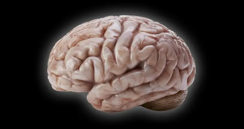
While most primates boast brains characterized by intricate folds and grooves, the common marmoset ( Callithrix jacchus) stands as a remarkable exception. These diminutive South American monkeys possess brains that are strikingly smooth, exhibiting minimal surface folding. This divergence from the typical primate cranial architecture has long intrigued neuroscientists, prompting a quest to understand the underlying developmental mechanisms. Now, researchers at the German Primate Center (DPZ), a Leibniz Institute for Primate Research in Göttingen, have made significant strides, identifying a cascade of cellular processes that illuminate the evolutionary path to the marmoset’s smooth brain. Their groundbreaking findings, published in the prestigious journal Science Advances, not only deepen our understanding of primate brain evolution but also hold potential implications for unraveling the complexities of human neurodevelopmental disorders.
Unveiling the Cellular Blueprint of Brain Development
The research team, led by scientists from the DPZ’s Junior Research Group Brain Development and Evolution, employed sophisticated miniature brain structures known as organoids. These three-dimensional cellular models, meticulously cultivated from human and common marmoset cells, serve as invaluable proxies for studying early brain development in vitro. By meticulously comparing these organoids, the scientists were able to pinpoint crucial differences in the behavior and development of neural progenitor cells – the crucial precursors responsible for generating neurons.
These progenitor cells are the architects of the developing brain, their proliferation rate and differentiation pathways dictating the ultimate size, complexity, and surface morphology of the cerebral cortex. In species with highly folded brains, such as humans, these progenitor cells undergo rapid and extensive division, generating a vast number of neurons. This sheer volume of neural tissue then necessitates an expansion of the cortical surface, leading to the characteristic folds and grooves that maximize surface area for enhanced cognitive function.
The marmoset, however, appears to have evolved a distinct strategy. The DPZ researchers discovered that several key differences in neural progenitor cell behavior converge to limit neuron production and, consequently, brain folding.
The Trio of Developmental Differences
The study identified three primary cellular mechanisms contributing to the marmoset’s smooth brain:
- Slower Progenitor Cell Division: A critical finding was that certain types of neural progenitor cells in common marmosets divide at a significantly slower pace compared to their human counterparts. This reduced rate of proliferation directly curtails the overall number of neurons generated during the critical developmental window.
- Simplified Cell Structure: Other progenitor cells in marmosets were observed to possess a simpler cellular structure, characterized by fewer branching processes. These less complex cellular architectures are less conducive to rapid proliferation, further contributing to a reduced output of new neurons.
- Compressed Developmental Window: Perhaps one of the most significant revelations was the difference in the temporal dynamics of progenitor cell activity. Marmosets exhibit a considerably shorter period during which these cells can undergo rapid multiplication and expansion. This compressed window of opportunity for rapid growth limits the total number of neurons that can be produced before the developmental stage concludes.
"We wanted to understand which changes at the cellular level cause the marmoset brain to grow less and form fewer folds," explains Lidiia Tynianskaia, one of the study’s two first authors and a PhD student in the Junior Research Group Brain Development and Evolution at the DPZ. "At the beginning of development, the common marmoset brain exhibits the typical structure and composition of a large, folded primate brain. As development progresses, processes must therefore occur that effectively slow down the production of nerve cells."
César Mateo Bastidas Betancourt, also a first author of the study and a PhD student in the Junior Research Group, elaborated on the findings: "Our investigations have shown that certain progenitor cells in the common marmoset divide significantly more slowly than in humans. Other progenitor cells have a simpler structure than their human counterparts, with fewer processes, and are therefore less proliferative. Both of these factors ultimately result in fewer nerve cells, which contributes to a smaller size and less folding of the cerebral cortex in marmosets."
The Power of Organoids in Unraveling Evolutionary Puzzles
The reliance on brain organoids proved instrumental in this research. These lab-grown miniature brains, while simplified representations, effectively recapitulate key aspects of early human and primate brain development. They offer a powerful platform for high-throughput experimentation and allow researchers to gather statistically robust data that would be prohibitively difficult or impossible to obtain from live primate embryos.
"The study combines the advantages of in vivo and in vitro methods," says Michael Heide, head of the Junior Research Group Brain Development and Evolution. "Organoids are well-suited for obtaining statistically robust results because such sample sizes are not feasible in primates. We subsequently repeated some key experiments in fetal brain tissue to confirm the results from the organoids."
A crucial aspect of the organoid methodology involved establishing a reliable correlation between the developmental stages observed in the organoids and those in natural marmoset brains. The researchers determined that a marmoset brain organoid cultured for approximately 50 days closely mirrors the developmental stage of a natural marmoset brain at around 90 days of gestation. This critical calibration allowed for meaningful comparisons and accurate interpretation of the experimental findings, bridging the gap between in vitro models and in vivo reality.
A Glimpse into the Evolutionary Landscape of Primate Brains
The common ancestor of modern primates is believed to have possessed a moderately sized brain with at least some degree of cortical folding. Over millions of years of evolution, diverse pressures and genetic adaptations have led to the vast array of primate brain sizes and complexities we observe today. The common marmoset, with its smooth brain, represents a fascinating evolutionary trajectory that deviates from the trend of increasing cortical folding seen in many other primate lineages, including that of humans.
The DPZ findings suggest that relatively subtle changes at the cellular and temporal levels of brain development can translate into significant differences in brain morphology. This underscores the principle that evolutionary innovation can arise from modifications in fundamental developmental processes rather than requiring entirely novel genetic blueprints. The research provides a compelling hypothesis for how the diverse spectrum of primate brain shapes and sizes emerged, explaining why some species retained or amplified cortical folding for enhanced cognitive abilities, while others, like the marmoset, evolved a different, yet effective, neural architecture.
Broader Implications for Human Health and Neuroscience
Beyond their evolutionary significance, these discoveries hold considerable promise for advancing our understanding of human neurodevelopmental disorders. Conditions such as microcephaly, lissencephaly (smooth brain disease), and certain forms of intellectual disability are characterized by abnormal brain size and folding. By elucidating the cellular mechanisms that govern normal primate brain development, scientists can gain critical insights into what goes awry in these conditions.
A deeper comprehension of the molecular and cellular pathways that regulate neuron production, progenitor cell proliferation, and the timing of developmental events could pave the way for novel diagnostic tools and therapeutic interventions. If researchers can identify specific genes or cellular processes that are dysregulated in smooth-brained primates, they may be able to apply this knowledge to understand and potentially treat similar conditions in humans.
The common marmoset’s unique brain structure, once a puzzle, has now become a vital key to unlocking fundamental questions about brain evolution and development. This research exemplifies the power of comparative studies and advanced model systems in pushing the boundaries of scientific knowledge, offering hope for future breakthroughs in both evolutionary biology and human medicine. The meticulous work of the DPZ scientists has not only illuminated the past but also illuminated a path forward for addressing some of the most challenging questions in neuroscience.

0 Comments